A fertility specialist orders a “saline infusion sonography” and the woman leaves the clinic with a form that has three words on it and no explanation. She has already had a transvaginal scan. She may have already had an HSG. Why does she need another test that also looks at the uterus?
I’m Dr. Rajashree NS, a consultant radiologist, and SIS is one of the tests I perform most often for women in the middle of a fertility workup, particularly after a failed embryo transfer or when a routine scan raises a question that needs a closer look. This guide walks through what the test actually does, how it differs from the scans you may have already had, what a normal and an abnormal result look like, and what typically happens next.
What this post covers:
- What saline infusion sonography is and how the procedure works
- Why it sees things a routine ultrasound and an HSG can miss
- What it finds: polyps, submucosal fibroids, adhesions, cavity shape
- Cycle timing, discomfort, and what to expect during the test
- Cost in India and what typically happens after the result
What Saline Infusion Sonography Is
Saline infusion sonography, also called sonohysterography or SHG, is a transvaginal ultrasound performed while a small amount of sterile saline is gently instilled into the uterine cavity through a thin catheter. The saline separates the front and back walls of the uterus, which are normally touching, so the cavity opens up like a room with the lights turned on.
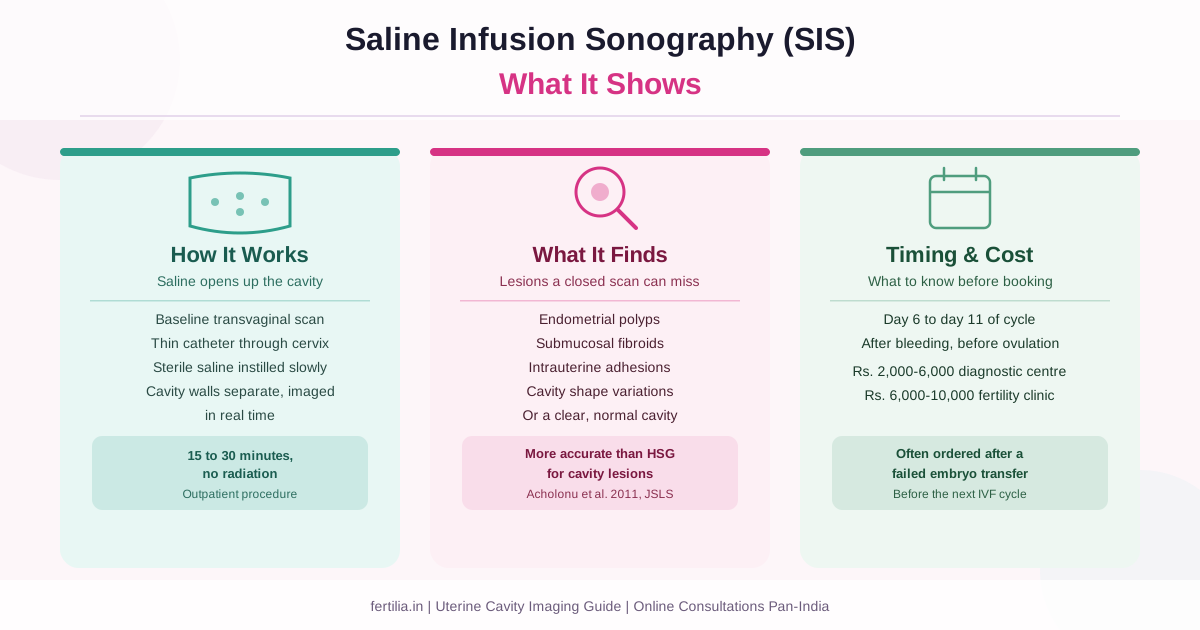
The procedure itself has two steps. First, a baseline transvaginal scan is done to look at the uterus and ovaries in their resting state, the same as any pelvic ultrasound. Then a narrow catheter is passed gently through the cervix, and sterile saline is slowly infused while the ultrasound probe images the cavity in real time. The saline fills the potential space between the uterine walls and outlines anything sitting inside it.
This is the detail that makes SIS different from every scan that came before it. A closed uterine cavity, imaged on a routine transvaginal scan, can hide a small polyp or a flat adhesion because the two walls are pressed together around it. Distend the cavity with fluid, and that same lesion stands out clearly against the fluid on either side of it.
The test typically takes 15 to 30 minutes from start to finish, including the baseline scan. The saline infusion itself is usually the shortest part, often five to ten minutes.
Why SIS Sees What Other Scans Miss
Three tests commonly get compared here, and each answers a different question.
Transvaginal scan (TVS) images the uterus and ovaries with the cavity in its normal, closed state. Our guide on what a transvaginal scan shows covers this in detail. A TVS is excellent for measuring the uterus, looking at the ovaries, and picking up larger fibroids or an obviously abnormal lining, but a small polyp sitting flush against the cavity wall can be genuinely difficult to see when the walls are collapsed against each other.
HSG (hysterosalpingogram) uses X-ray and a contrast dye instead of ultrasound, and its main strength is confirming whether the fallopian tubes are open, something SIS does not routinely assess. Our guide on how to read your HSG report explains that test in full. For the cavity itself, though, HSG has real limits. A comparison study using hysteroscopy as the reference standard found sonohysterography had sensitivity of 81.8 percent and specificity of 93.8 percent for intrauterine abnormalities, against 58.2 percent and 25.6 percent for HSG (Acholonu UC et al., JSLS, 2011). In plain terms, HSG both misses more real cavity problems and flags more shadows that turn out not to be anything, while SIS is considerably more reliable for exactly this question.
SIS fills the gap between the two. It gives a detailed look at the cavity itself, closer to what HSG offers for the tubes, without radiation or contrast dye, and it is generally more accurate than either a plain TVS or an HSG for identifying focal cavity lesions.
None of the three tests replaces the others outright. A woman may reasonably have all three across a fertility workup, each answering a different part of the question.
What SIS Finds
Endometrial polyps. A polyp typically appears as a smooth, rounded lesion outlined by fluid on all sides, sometimes with a single feeding blood vessel visible on Doppler. This is the finding SIS is particularly good at picking up, because a flat or broad-based polyp is one of the easiest lesions to miss on a routine closed-cavity scan. Our guide on reading a fibroid ultrasound report covers the related but distinct finding of fibroids on imaging.
Submucosal fibroids. These are fibroids that bulge into the cavity rather than staying within the muscular wall. SIS shows how much of the fibroid projects into the cavity and how much it distorts the shape of the uterine lining, which matters for deciding whether hysteroscopic removal is appropriate before a fertility treatment. The fibroids and natural conception guide explains how location, more than size alone, determines fertility impact.
Intrauterine adhesions. Also called Asherman’s syndrome, adhesions are bands of scar tissue, usually from a previous D&C or infection, that can bridge across the cavity or prevent it from distending fully with saline. When the cavity does not fill evenly, or a band is visible crossing it, this is a specific finding that changes management. Our Asherman’s syndrome guide covers diagnosis and treatment in more depth.
Cavity shape. Some women have a naturally different uterine shape, such as a septum, that only becomes clearly visible once the cavity is filled with fluid and imaged from multiple angles. This is a separate finding from the ones above and usually needs its own conversation about what, if anything, needs to be done.
A normal result. In many women, SIS shows a smooth, regularly shaped cavity that fills evenly with saline and has no lesion at all. This is a genuinely reassuring finding, not an inconclusive one, and it rules out the cavity as the reason for an unexplained symptom or a failed transfer.
If your report has already come back with one of these findings and you are not sure what it means for you specifically, a video consultation is the fastest way to get a clear answer.
Dr. Suganya Venkat is available online, pan-India, for a Rs. 399 video consultation. You can share your scan report directly in the conversation.
When SIS Is Ordered
SIS is not typically the first scan in a fertility workup. It is usually ordered for a specific reason:
Before starting IVF, when the cavity has not yet been assessed in detail, especially if a routine scan raised a question, there is a history of abnormal bleeding, or a prior D&C or uterine surgery makes adhesions a reasonable concern.
After a failed embryo transfer. A single failed transfer, even with a good embryo, is common and does not by itself point to a cavity problem. Repeated failures are a different picture, and reassessing the cavity is a standard part of that investigation. Our recurrent implantation failure guide covers where SIS fits in that broader sequence.
When symptoms suggest a cavity lesion. Irregular spotting between periods, unusually heavy bleeding, or bleeding after intercourse can all point toward a polyp or other focal lesion that a routine scan did not fully characterise.
As a follow-up to an unclear finding on a routine scan. If a transvaginal scan shows the endometrial lining as thickened, irregular, or difficult to assess clearly, SIS is often the next step before deciding whether hysteroscopy is needed.
If SIS finds something that needs to be removed or biopsied, such as a polyp or a submucosal fibroid, the next step is usually a hysteroscopy, which can both look directly at the cavity and treat the lesion in the same procedure. SIS is a diagnostic look; hysteroscopy is the tool that treats what SIS finds.
Cycle Timing, Preparation & What to Expect
SIS is scheduled after your period has fully ended but before ovulation, typically around day 6 to day 11 of the cycle, counting day 1 as the first day of full flow. This timing matters for two reasons: the lining is at its thinnest, which makes it easier to see a small lesion against it, and scheduling before ovulation avoids the small possibility of disturbing an early pregnancy.
You do not need to fast. A full bladder is not required the way it sometimes is for an abdominal scan, since this is done transvaginally. Some clinics suggest taking a simple pain reliever roughly 30 to 60 minutes before the appointment, though this varies by clinic and is worth confirming with whoever ordered your test, particularly if you have a reason to avoid NSAIDs.
Most women describe the sensation during saline infusion as period-like cramping, similar to what many feel during an IUD insertion, that settles quickly once the saline stops flowing. Reported experiences vary considerably from person to person, and cervical sensitivity, anxiety, and how quickly the fluid is instilled all play a role. Light spotting or watery discharge for a day afterward is common and expected.
When SIS is not done: if pregnancy is known or possible, if there are signs of an active pelvic infection such as fever or unusual discharge, or during heavy active bleeding, since blood in the cavity makes the images difficult to interpret. Contact your clinic if you develop fever, worsening pelvic pain, or foul-smelling discharge in the days after the test, though this is uncommon.
SIS Cost in India
Saline infusion sonography typically costs Rs. 2,000 to Rs. 6,000 at a diagnostic centre or radiology lab, rising to Rs. 6,000 to Rs. 10,000 at a private fertility clinic chain where the scan is bundled with a same-day consultation. As with any pelvic imaging test, government hospitals and medical colleges may offer it at a lower rate for OPD patients, similar to the pricing pattern for HSG.
Before booking, it is worth confirming exactly what the quoted price includes: the transvaginal ultrasound itself, the catheter and saline, the radiologist’s report, and whether a same-day written report is part of the fee or billed separately. “SIS” is sometimes used loosely by clinics to mean slightly different things, so if tubal assessment is also part of what your doctor wants checked, ask specifically whether that is included or whether it needs a separate test.
What Happens After the Result
A normal result means the cavity is clear, which is a genuinely useful finding: it rules out the uterine cavity as a cause of unexplained bleeding or a failed transfer, and your clinical team moves on to other parts of the investigation.
A polyp or submucosal fibroid usually leads to a hysteroscopy, both to confirm the finding directly and to remove it in the same sitting if appropriate. Removal before the next embryo transfer is standard practice when a cavity lesion is found in the context of fertility treatment.
Adhesions are also typically confirmed and treated by hysteroscopy, since SIS can suggest their presence but hysteroscopy allows direct visualisation and, often, treatment in the same procedure.
An unusual cavity shape is discussed on its own terms, since not every variation needs correction, and the decision depends on your fertility history and what you and your doctor are trying to achieve.
In every case, the SIS result is one piece of information that your gynaecologist or fertility specialist reads alongside your history, your symptoms, and any earlier scans, not a standalone verdict.
If you have an SIS report and are not sure what your specific result means for your next step, Dr. Suganya Venkat can walk through it with you directly.
Talk to Dr. Suganya Venkat over a video consultation, pan-India, at Fertilia.
Frequently Asked Questions
What is saline infusion sonography used for?
It is used to get a detailed look inside the uterine cavity, mainly to check for polyps, submucosal fibroids, and adhesions that a routine ultrasound can miss because the cavity walls are normally collapsed against each other. It is commonly ordered as part of a fertility workup, especially after a failed embryo transfer, or to investigate abnormal bleeding.
Is SIS the same as sonohysterography?
Yes. Saline infusion sonography, sonohysterography, and SHG all refer to the same procedure, an ultrasound performed while saline is gently instilled into the uterine cavity through a thin catheter.
Is SIS painful?
Most women describe the sensation as period-like cramping during the saline infusion, comparable to what many feel with an IUD insertion, that settles quickly once the infusion stops. How uncomfortable it feels varies from person to person, and your clinic can advise on taking a pain reliever beforehand if you are prone to cramping.
How is SIS different from HSG?
HSG uses X-ray and an iodine-based contrast dye and is primarily used to check whether the fallopian tubes are open. SIS uses ultrasound and saline and is primarily used to examine the uterine cavity in detail. A comparison study found SIS considerably more accurate than HSG for detecting cavity lesions like polyps and adhesions, while HSG remains the more established test for tubal patency. Many women have both tests as part of a complete fertility workup, since they answer different questions.
When in my cycle is SIS done?
Typically between day 6 and day 11 of the menstrual cycle, after bleeding has stopped but before ovulation. This timing keeps the endometrial lining thin, which makes small lesions easier to see, and avoids the small chance of an early pregnancy being present.
How much does saline infusion sonography cost in India?
Typically Rs. 2,000 to Rs. 6,000 at a diagnostic centre, and Rs. 6,000 to Rs. 10,000 at a private fertility clinic chain when bundled with a consultation. Always confirm what the quoted price includes before booking.
What happens if SIS finds a polyp or fibroid?
The usual next step is a hysteroscopy, which can directly view the cavity and remove the polyp or submucosal fibroid in the same procedure. This is generally recommended before the next embryo transfer if you are undergoing fertility treatment.
An SIS report with unfamiliar findings does not need to stay a mystery. If you have a report you would like explained, or your doctor has recommended the test and you want to understand why, Dr. Suganya Venkat is available online for a Rs. 399 video consultation, pan-India, at Fertilia. Start the conversation on WhatsApp.
For the complete guide to fertility testing and getting pregnant after diagnosis, download the free Getting Pregnant guide.
Dr. Rajashree NS is a Consultant Radiologist with an MD in Radio-diagnosis from Sree Mookambika Institute of Medical Sciences. She interprets pelvic and fertility ultrasound reports as part of the Fertilia Health team.