A woman comes to the radiology suite holding a prescription. The sheet says “TVS” or “transvaginal ultrasound.” Nobody has told her what to expect. She has read a few things online and is not sure what is about to happen, whether it will hurt, or why her doctor did not just ask for a normal scan of her abdomen.
That uncertainty is extremely common. The transvaginal scan is one of the most frequently ordered pelvic imaging tests in gynaecology, fertility work, and early pregnancy, yet the preparation instructions are often minimal and the reason for choosing it over an abdominal scan is rarely explained.
I am a consultant radiologist, and I do this scan routinely. This guide covers what a TVS is, why it produces better images than an abdominal scan for most pelvic structures, what it actually shows us, how to prepare, and what the experience is like. If your doctor has ordered a TVS, reading this first should answer most of the questions you are carrying to the appointment.
What this post covers:
- What a transvaginal scan is and how the probe works
- Why TVS rather than an abdominal ultrasound
- What the scan images: ovaries, uterus, lining, follicles, early pregnancy
- How to prepare (bladder, clothing, timing)
- What the experience is like, and honest reassurance on discomfort
- What TVS is ordered for, and how it fits into the larger scan-interpretation cluster
- FAQ
What a Transvaginal Scan Is
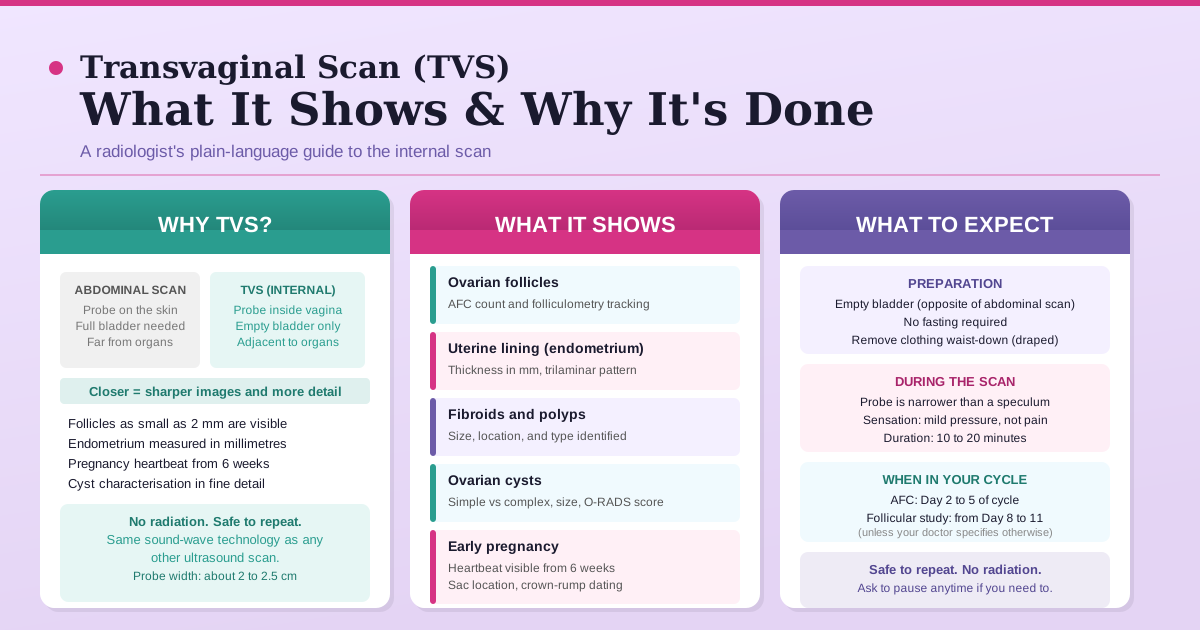
A transvaginal scan is an ultrasound examination in which a narrow probe, covered with a gel-coated sheath, is gently placed into the vaginal canal to image the pelvic organs from the inside. It uses the same sound-wave technology as every other ultrasound: no X-ray, no radiation, nothing injected.
The probe itself is typically about 2 to 2.5 cm wide at its widest point, which is narrower than a standard gynaecological speculum. It is placed only a few centimetres inside the vagina. The radiologist or sonographer manoeuvres it gently to view the uterus and both ovaries from different angles.
The scan takes roughly 10 to 20 minutes depending on what is being assessed. You remain clothed from the waist up and are draped throughout for privacy.
Why TVS Gives Better Images Than an Abdominal Scan
The abdominal ultrasound probe sits on the skin above the abdomen and sends sound waves through layers of skin, fat, muscle, bowel gas, and bladder fluid before they reach the uterus and ovaries. Distance and intervening tissue reduce clarity.
The transvaginal probe is placed directly adjacent to the uterus, ovaries, and fallopian tubes, with almost nothing in between. This proximity produces images that are significantly sharper and more detailed.
This matters clinically for several reasons:
Follicle counting and sizing. Small follicles, including the 2 to 10 mm antral follicles counted during an antral follicle count (AFC), are reliably visible only on TVS. An abdominal scan may miss small follicles or undercount them, which would give a falsely low ovarian reserve picture.
Endometrial lining. The uterine lining (endometrium) needs to be measured in millimetres, and the trilaminar pattern that matters for fertility assessment is only clearly visible at high resolution. Endometrial thickness assessment is almost always done transvaginally for this reason.
Early pregnancy. A fetal heartbeat can be seen on TVS from around 6 to 7 weeks of pregnancy. On an abdominal scan, the same finding may not be visible until 10 to 11 weeks. For any early pregnancy scan where the precise number of weeks is uncertain, TVS is the standard choice.
Ovarian cysts and abnormalities. A small cyst or a subtle structural change in the ovary is far more reliably detected and characterised transvaginally. The ovarian cyst scan report your radiologist writes draws on TVS images for most of its detail.
An abdominal scan is still used in certain situations, including later pregnancy (second and third trimester) and some abdominal assessments, but for any pelvic evaluation in a non-pregnant woman or in early pregnancy, TVS is the standard of care.
What a TVS Shows
A single TVS appointment can provide information about several structures simultaneously. Here is what the radiologist is examining:
The Uterus
The scan records the uterus size (length, width, depth), shape, and position. It identifies the presence and approximate size of fibroids (solid growths in or on the uterine wall) or polyps (small projections from the inner lining). It also shows whether the cavity looks regular or distorted.
The Endometrium (Uterine Lining)
The lining thickness is measured in millimetres. The pattern (whether it appears trilaminar, uniform, or irregular) is noted. This measurement changes across the cycle and is relevant to fertility planning, response to ovulation induction medicines, and in some cases to ruling out pathology.
The Ovaries
Both ovaries are located and measured. The scan counts and sizes the follicles visible on each ovary, which is the basis of the AFC test used to estimate egg reserve. In a follicular study, the dominant follicle is tracked across multiple visits as it grows toward ovulation.
The ovaries are also examined for cysts, their appearance (simple or complex), vascularity on Doppler, and structural changes consistent with PCOS (polycystic ovarian morphology on ultrasound is characterised by a specific count and arrangement of small peripheral follicles, assessed using Rotterdam Criteria, 2003).
Early Pregnancy
A TVS in early pregnancy checks for the pregnancy sac, its location inside the uterus (to rule out an ectopic), the developing embryo, and the presence of a heartbeat. It also dates the pregnancy by measuring crown-rump length (CRL), which in the first trimester gives the most accurate gestational age estimate.
The Fallopian Tubes
The tubes themselves are not usually visible on a routine TVS unless they are enlarged or fluid-filled. If a tube is distended with fluid (a hydrosalpinx), the TVS will pick it up. Assessment of tubal patency requires a separate procedure, the HSG or hystero-salpingo-graphy, which is different from a standard TVS.
When a TVS Is Ordered
Your doctor will request a TVS in a range of clinical situations. The most common ones:
- Fertility evaluation. To count antral follicles (AFC), assess the endometrial lining, and check the ovaries and uterus before or during fertility treatment.
- Follicular monitoring (folliculometry). To track the growing dominant follicle across a cycle and time ovulation or IUI.
- Irregular periods or PCOS evaluation. To count ovarian follicles and assess uterine structure.
- Early pregnancy. To confirm the pregnancy is inside the uterus, measure the heartbeat, and date the pregnancy.
- Suspected ovarian cyst. To characterise whether a cyst is simple, complex, or needs further evaluation.
- Pelvic pain or abnormal bleeding. To rule out structural causes such as fibroids, polyps, or ovarian pathology.
- Endometrial assessment. In women with abnormal bleeding, to assess lining thickness and uniformity.
If your prescription says “TVS” for any of these indications, the test is appropriate and well-matched to what needs to be looked at.
How to Prepare for a TVS
The preparation for a TVS is the opposite of an abdominal pelvic scan.
Bladder: empty, not full. For an abdominal scan, a full bladder is required because it pushes the bowel out of the way and creates an acoustic window through which the probe can image the uterus. For a TVS, the probe is already inside the pelvic cavity, so the bladder just needs to be comfortably empty. Arriving with a very full bladder is actually uncomfortable during TVS. Use the bathroom before the scan.
Timing in the cycle. If your doctor has specified a particular day of your cycle, follow that instruction. An AFC is done on Day 2 to 5. A follicular study begins around Day 8 to 11. For other indications (cyst assessment, early pregnancy, structural evaluation), the day of the cycle usually does not matter.
Clothing. You will be asked to remove clothing from the waist down and will be given a drape. There is no other special clothing preparation needed.
No fasting. Food and water intake before the scan makes no difference to TVS image quality.
During your period. A TVS can be done during menstruation if the clinical indication requires it (for example, an early cycle AFC). It is your choice; some women prefer to wait, and that is fine if it does not delay necessary clinical decisions.
If your doctor has not specified a particular day and the scan is for general assessment, booking it on any comfortable day of your cycle is acceptable.
Have questions about your TVS report? Speak with Dr. Rajashree via our online consultation. →
Is a TVS Safe? Is It Painful?
Safety. A transvaginal scan is completely safe. It uses ultrasound sound waves, not X-rays. There is no radiation. The probe does not enter the uterus or pass the cervix. Sonographic imaging in this context has been used for decades and is the standard method for pelvic assessment in women worldwide.
It is safe during early pregnancy. It is safe to repeat as often as clinically needed, which is why follicular monitoring involves several scans across a single cycle without concern.
Discomfort. Most women describe the sensation as mild pressure, similar to the feeling of a full bladder, that comes and goes as the probe is angled to view each structure. A few women find one or two positions mildly uncomfortable, particularly if there is existing pelvic tenderness from a cyst or endometriosis. The word “painful” is not accurate for most women; “pressure” or “a little odd” is much closer to the typical experience.
If at any point during the scan you feel genuinely uncomfortable, tell the radiologist or sonographer. The probe can be repositioned or the angle adjusted. You can also ask for a brief pause if needed.
Virginity and unmarried women. The TVS probe is narrow but it does enter the vaginal canal. If you have not had penetrative sex, please inform your doctor and the radiology team before the scan. In these situations, a transabdominal approach with a very full bladder can often provide sufficient information, or the TVS can be discussed with a modified technique. The clinical team will take your preference and situation into account.
Reading Your TVS Report: What the Language Means
A standard TVS report uses technical phrasing that makes sense to the requesting doctor but can read as a wall of unfamiliar terms. A few of the most common ones:
“Uterus anteverted / retroverted” refers to which way the uterus tilts. Anteverted (tilting forward) is more common. Retroverted (tilting backward) is a normal variation in roughly 20% of women and rarely affects fertility or health.
“Endometrial thickness X mm, echogenic / trilaminar” gives the lining measurement and its texture pattern. Trilaminar (three-layered) is the pattern seen in the mid-proliferative phase and is considered a good sign for receptivity.
“Right ovary: X × Y mm, X follicles seen” is the ovary measurement and follicle count. For an AFC report, this count on both sides together is the antral follicle count.
“Adnexal cyst / anechoic area / simple cyst” describes a fluid-filled structure. “Anechoic” means no internal echoes (clear fluid), which is associated with simple, likely benign cysts. For more detailed guidance on cyst terminology, the ovarian cyst scan report guide covers this in full.
“No free fluid in the pouch of Douglas” refers to the space behind the uterus. A small amount of free fluid after ovulation is completely normal. Larger amounts or fluid with an unusual appearance may warrant further evaluation.
If a term on your report is not listed here, bring the printout to your consultation. Your treating doctor is the right person to interpret any finding in the context of your complete clinical picture.
The TVS in Your Scan Journey
The transvaginal scan is often the first imaging step in a fertility or gynaecology workup, and it connects to several other scans and tests you may encounter:
- AFC (done on Day 2 to 5 by TVS) estimates your egg reserve. See the AFC guide for how the count is interpreted by age.
- Folliculometry is a series of TVS scans that track your dominant follicle across one cycle. See the follicular study guide for what the report numbers mean.
- HSG is a separate procedure for checking whether the fallopian tubes are open. It is not a TVS. The HSG report guide explains that test separately.
Understanding which scan does what, and why your doctor has ordered a specific one, helps you arrive at each appointment prepared rather than anxious.
Questions about your scan report? Reach us online for a report interpretation consultation. →
Frequently Asked Questions
What is the difference between a transvaginal scan and an abdominal ultrasound? An abdominal ultrasound uses a probe on the skin above the belly and requires a full bladder. A transvaginal scan uses a narrow probe placed gently inside the vaginal canal and requires an empty bladder. TVS provides significantly sharper images of the uterus and ovaries because the probe is much closer to those structures, with minimal tissue between the probe and the organs being imaged.
Is a transvaginal scan painful? For most women, the sensation is mild pressure rather than pain, similar to the feeling of a moderately full bladder. The probe is narrower than a gynaecological speculum. If you have existing pelvic tenderness (from endometriosis or an inflamed cyst, for example), you may feel more discomfort in specific positions. Always let the radiologist or sonographer know if you are uncomfortable; the probe angle can be adjusted and a brief pause is always possible.
Is a transvaginal scan safe? Does it use radiation? A TVS uses sound waves, not X-rays, so there is no radiation involved. It is safe for all women including those in early pregnancy. The probe does not enter the uterus or cervix. The scan can be repeated as many times as clinically needed (follicular monitoring, for example, involves multiple scans in one cycle) without any safety concern.
How should I prepare for a transvaginal scan? Do I need a full bladder? Unlike an abdominal pelvic scan, a TVS requires an empty bladder. Arrive having used the bathroom beforehand. There is no fasting required. If your doctor has specified a particular day of your cycle, follow that instruction; for other indications, any comfortable cycle day is fine.
Why is TVS used in early pregnancy instead of a regular abdominal scan? In the first six to eight weeks of pregnancy, the uterus sits deep in the pelvis and is small. The abdominal scan’s distance from the organ means the embryo may not yet be visible through that route. A TVS shows the pregnancy sac and heartbeat reliably from around 6 weeks. It also confirms the pregnancy is located inside the uterus rather than in a fallopian tube (ectopic pregnancy), which is important to rule out early.
Can a TVS be done during my period? Yes, it can be done safely during menstruation. The most common reason to schedule a TVS specifically during a period is the antral follicle count, which should be done on Day 2, 3, 4, or 5 of the cycle. For other indications, menstruation does not affect the scan’s safety or the image quality in a way that matters clinically. If you prefer not to have the scan during your period and the timing is flexible, it is fine to reschedule.
Internal scan karana chahiye kab? (When should I have an internal scan?) Your doctor will specify the right time based on the reason for the scan. For an antral follicle count (AFC), it should be done on Day 2 to 5 of your cycle (counting the first day of bleeding as Day 1). For follicular monitoring, scans begin around Day 8 to 11. For early pregnancy confirmation, it is done as soon as pregnancy is suspected or confirmed. For ovarian cysts, fibroids, or general pelvic assessment, any comfortable day of the cycle is suitable unless your doctor specifies otherwise.
What to Take Away
A transvaginal scan is a standard, safe, non-radiation imaging procedure that gives your clinical team a detailed view of the uterus, uterine lining, ovaries, and follicles that an abdominal scan simply cannot match for resolution. Preparation is minimal: an empty bladder and any day-of-cycle instruction your doctor has given. The probe is narrow, the procedure is brief, and the discomfort for most women is mild pressure rather than pain.
If your doctor has ordered a TVS, it is because the pelvic structures under question are best seen this way. The information it provides connects directly to the clinical decisions that follow, whether that is timing treatment, tracking ovulation, characterising a cyst, or confirming an early pregnancy.
Arrive knowing what to expect, and the appointment itself is very straightforward.