Your HSG report may arrive before you can speak to the doctor who requested it. Words such as “cornual block” and “loculated spill” need explanation, but a report alone cannot tell you whether you need IVF.
The quick translation:
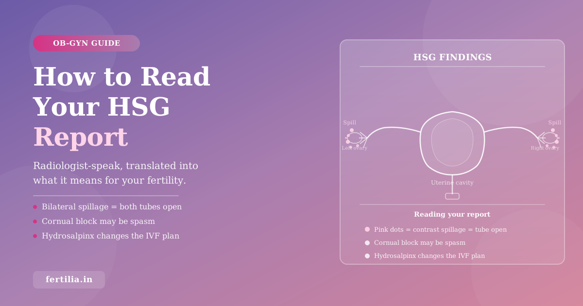
- Bilateral free spillage: both tubes are open to dye. It does not prove every aspect of tubal function is normal.
- Unilateral spillage: one tube is confirmed open. Natural conception may still be possible.
- Cornual block: an apparent blockage near the uterus. Spasm or debris can mimic a structural obstruction.
- Hydrosalpinx: a fluid-filled, damaged tube. Discuss its management before IVF.
- Filling defect: an area inside the womb needs a closer look, usually with saline ultrasound or hysteroscopy.
I’m Dr. Suganya Venkat, an OB-GYN with 15+ years of experience. This guide explains the report terms and the questions to take to your referring doctor.
What an HSG Is
An HSG (hysterosalpingogram) is an outpatient X-ray procedure done in the radiology department of a hospital or imaging centre. A thin catheter is passed through the cervix into the uterine cavity. A small volume of radio-opaque contrast dye is injected, and its spread through the uterus and tubes is observed under X-ray fluoroscopy in real time.
The test shows two things: the internal shape of the uterine cavity, and whether each fallopian tube is open. If the tubes are open, the dye flows through their entire length and spills into the pelvic cavity on each side, appearing as a “spillage” pattern on the X-ray image. If a tube is blocked, the dye cannot pass and the obstruction point is visible.
HSG is scheduled after menstrual bleeding has ended and before ovulation. The centre should confirm the date for your cycle and how to exclude pregnancy; calendar timing alone cannot exclude it. RadiologyInfo’s HSG patient guide, reviewed in 2024, describes preparation and safety precautions.
The test is useful but imperfect. Swart et al.’s meta-analysis in Fertility and Sterility (1995, PMID 7641899) reported sensitivity of 65 percent and specificity of 83 percent for tubal obstruction in the subset of studies where HSG and laparoscopy were assessed independently. These are study-level accuracy estimates, not your personal probability of having a blocked tube. HSG can miss an obstruction or suggest one that is not permanent.
This is Layer 7 of the fertility workup covered in The Honest Fertility Workup: An OB-GYN’s Indian Guide. If you have not seen the full workup picture, that post is the right starting point.
The Anatomy of an HSG Report
Indian HSG reports from radiology departments and imaging centres follow a broadly consistent structure. Understanding the layout helps you find the relevant information quickly.
A typical Indian HSG report has four parts:
Patient details and procedure note. Your name, age, cycle day, and a brief sentence confirming the procedure was performed with a specified type of contrast dye (usually a water-soluble contrast such as iohexol or iopromide).
Uterine cavity findings. The first radiological observation: the size, shape, and contour of the uterine cavity, and whether any filling defects are visible inside it.
Fallopian tube findings. Whether each tube filled with contrast and whether the contrast “spilled” at the fimbrial end. Any specific observations about tube calibre, regularity, or appearance.
Impression (or Conclusion). The radiologist’s summary, usually one or two lines of compressed clinical shorthand. This is what most women read first, and it almost always needs unpacking.
Decoding the Uterine Cavity Section
The uterine cavity section tells you about the internal shape of your uterus. Here are the phrases you are most likely to encounter:
“Normal uterine cavity outline” or “uterine cavity appears normal”: The dye outlines an expected cavity shape. This is reassuring, but HSG does not assess every condition in the uterine wall or reliably exclude small cavity lesions.
“Filling defect”: Something inside the cavity is preventing the dye from filling that region fully. The dye shows the outline of the obstruction. This is a descriptive observation, not a diagnosis. Filling defects most commonly represent submucosal fibroids, endometrial polyps, or intrauterine adhesions. Your next step is a hysteroscopy or saline infusion sonography to characterise exactly what it is and where.
“Septate uterus”: The cavity appears divided by a band of tissue. Before considering treatment, the diagnosis needs confirmation with imaging that also shows the outside of the uterus.
“Bicornuate uterus”: A congenital shape variation with two uterine horns. HSG cannot reliably distinguish this from a septate uterus because it shows the cavity but not the external uterine contour. Three-dimensional ultrasound or MRI may clarify the finding. The treatment decisions differ, so neither label should lead straight to surgery on an HSG result alone. This limitation is explained in ASRM’s Fertility evaluation of infertile women, 2021.
“Arcuate uterus”: A mild indentation at the top (fundus) of the uterine cavity. This is a normal variant. It does not require treatment.
“Adhesions” or “Asherman’s syndrome”: Bands of scar tissue inside the uterine cavity, typically following a curettage procedure (D and C), repeated infections, or prior uterine surgery. On HSG these appear as irregular filling defects or a distorted cavity outline. Hysteroscopic adhesiolysis is the treatment.
“Intrauterine polyp” or “submucosal fibroid”: Both create filling defects. A polyp is a soft tissue overgrowth from the endometrium. A submucosal fibroid protrudes into the cavity from the uterine wall. Both can be confirmed and removed hysteroscopically.
| Finding | What it means | Next step |
|---|---|---|
| Normal cavity outline | No cavity abnormality visible on this test | Read alongside ultrasound and symptoms |
| Filling defect | Something inside the cavity | Hysteroscopy or saline sonography |
| Suspected septate uterus | Possible midline division | Confirm with 3D ultrasound or MRI |
| Suspected bicornuate uterus | Possible two-horned uterus | Confirm external uterine shape |
| Arcuate uterus | Mild fundal dip | No treatment needed |
| Adhesions | Scar tissue inside cavity | Hysteroscopic adhesiolysis |
| Polyp or submucosal fibroid | Growth inside cavity | Hysteroscopic removal |
Decoding the Fallopian Tubes Section
This is the section most women turn to first, and the one with the most consequential language. I will go through every phrase you are likely to see, in order of what they mean clinically.
“Bilateral spillage seen” or “bilateral free spillage”: Both fallopian tubes filled completely with contrast and the dye spilled freely into the pelvic cavity on both sides. Both tubes are patent (open). That is reassuring, but it does not assess the microscopic lining of the tubes or rule out surrounding adhesions.
“Unilateral spillage” or “spillage seen on the left only” (or right only): One tube has confirmed free spillage and the other does not. With one healthy open tube, natural conception remains possible. An egg can sometimes be picked up from the opposite ovary; this has been documented after removal of one tube (Ross et al., Human Reproduction, 2013, PMID 23381619). It does not mean every opposite-side ovulation will lead to pregnancy. For the full clinical picture of what this diagnosis means, including treatment options, see Blocked Fallopian Tubes: Causes, Signs and Fertility Impact.
“No spillage seen bilaterally” or “bilateral tubal occlusion”: Neither tube showed dye exiting into the pelvis. The location of the apparent obstruction matters. If both appear blocked near the uterus, further assessment is needed to exclude spasm or a technical problem before treating the finding as permanent. The repeat-HSG study below explains why. Confirmed blockage of both tubes changes the conception plan, but the report alone does not choose between further testing, tubal treatment and IVF.
“Cornual block”, “proximal block”, or “interstitial block”: The apparent obstruction is at the cornua, the point where the tube enters the uterine wall. Temporary muscle contraction, mucus or debris can prevent dye passage and mimic a fixed blockage. If this result would change your treatment, ask your doctor whether it needs confirmation before deciding on surgery or IVF. (The full explanation of spasm versus structural block is in the next section.)
“Mid-tubal block” or “isthmic block”: A blockage in the middle segment of the tube, between the cornua and the fimbrial end. Your doctor will assess whether this fits prior infection, surgery or another cause of scarring. The report location alone cannot establish the cause. Treatment depends on the extent of damage and the rest of your fertility assessment.
“Distal block” or “fimbrial block”: A blockage at the ovarian end of the tube, where the fimbriae (the finger-like projections that sweep in the released egg) are located. The fimbriae are the most delicate part of the tube and the most easily damaged by infection and endometriosis.
“Hydrosalpinx”: A specific finding where a distal blockage has caused fluid to accumulate inside the tube, creating a swollen, fluid-filled segment. This is clinically significant beyond the blockage itself. The fluid is associated with poorer implantation. Backward flow into the uterus, reduced endometrial receptivity and a possible direct effect on embryos have been proposed; the exact mechanism is not settled. ASRM’s Role of tubal surgery in the era of assisted reproductive technology (2021, PMID 33642065) supports discussing salpingectomy (removal) or proximal occlusion (blocking the tube’s connection to the uterus) before IVF for communicating hydrosalpinges. Repair may be considered in selected mild cases. The plan depends on the tube’s condition, surgical risks and whether you are trying naturally or having IVF.
“Loculated contrast” or “pelvic loculation”: The contrast dye, after spilling from the tube, pooled in pockets rather than spreading freely through the pelvic cavity. This suggests adhesions around the tube, which may follow infection, endometriosis or surgery. If dye has exited the tube, that demonstrates passage through it; the concern is how it spreads afterward. Loculation suggests rather than proves adhesions. Your doctor will decide whether symptoms and the full fertility assessment justify laparoscopy, following ASRM’s 2021 fertility-evaluation guidance.
“Beaded appearance” or “irregular tubes with beading”: A pattern where the tube appears segmentally dilated and irregular rather than smooth. Genital tuberculosis is one possible cause worth considering in India, but an HSG pattern cannot diagnose active TB. Assessment may include sampling for culture, molecular testing and histology, chosen by your gynaecologist and TB specialist. A Mantoux test alone cannot diagnose active genital TB, and a negative result does not exclude it. See Tzelios et al., Open Forum Infectious Diseases (2022, PMID 36447614).
“Contrast extravasation”: Dye is described outside its intended path. Ask the radiologist what happened and whether the study still assessed both tubes adequately; do not assume this is ordinary free spillage.
| Report phrase | What it means | What to do next |
|---|---|---|
| Bilateral spillage seen | Both tubes open to dye | Review the other fertility factors |
| Unilateral spillage | One tube confirmed open | Clarify the other tube’s finding |
| No spillage bilaterally | Neither tube showed dye exiting | Confirm proximal findings before treatment decisions |
| Cornual block | Apparent block at uterus-tube junction | Discuss confirmation if it changes treatment |
| Mid-tubal block | Apparent obstruction in tube middle | Assess cause and extent of damage |
| Distal block or fimbrial block | Obstruction at ovarian end | Assess anatomy and look for hydrosalpinx |
| Hydrosalpinx | Fluid-filled damaged tube | Discuss management before IVF |
| Pelvic loculation | Dye pockets suggest adhesions | Consider further assessment if indicated |
| Beaded tubes | Irregular tube contour | Consider causes including TB; not a diagnosis |
💜 Unsure what your specific HSG result means for your situation? Message Dr. Suganya’s team on WhatsApp and share your findings. We will walk you through it before your next appointment.
What “Spasm vs True Block” Means
Muscle at the uterus-tube junction can contract during dye injection, preventing contrast from passing. Mucus or debris can also obstruct the opening. A single image cannot reliably separate these from fixed scarring, and this is not something you caused by being anxious.
There is direct evidence for checking an apparent proximal block. In Dessole et al.’s prospective study, 24 of 40 women with proximal obstruction on their first HSG had both tubes patent on a repeat about one month later (Fertility and Sterility, 2000, PMID 10785234). That is a repeat-test finding, not proof that all the original results were spasm.
Your doctor may discuss:
- Repeat HSG: useful when the first study was technically limited or a proximal result needs clarification.
- Ultrasound contrast testing (HyCoSy or HyFoSy): assesses dye or foam passage without X-rays. A basic saline cavity scan is not equivalent to a test that documents each tube separately.
- Selective salpingography or tubal cannulation: assesses the opening of the affected tube and can sometimes clear an obstruction.
- Laparoscopy with dye testing: can inspect the tubes and surrounding pelvis when surgery is otherwise indicated. It is invasive, is not infallible, and is not required after every abnormal HSG.
ASRM’s 2021 tubal-surgery opinion also notes that isolated unilateral proximal obstruction without distal abnormalities does not always require intervention. Ask what a further test would change in your plan.
HSG Cost in India and What to Expect on the Day
Before booking at an Indian hospital or diagnostic centre, ask for a written total: does it include the catheter, contrast, radiologist’s report and any medicines? Check whether you will receive the images as well as the report. Bring both to the follow-up. Use the HSG cost guide to prepare your budgeting questions, then confirm the total directly with the centre.
Before the test: tell the centre if pregnancy is possible, if you have symptoms of pelvic infection or an untreated sexually transmitted infection, or if you have previously reacted to iodinated contrast. HSG should not be performed with possible pregnancy or active pelvic infection. A previous contrast reaction needs an individual safety plan. These precautions are set out in RadiologyInfo’s HSG guide.
Pain relief: ask the team what to take and when, rather than choosing a dose from an online guide. Oral painkillers are sometimes offered, but the Cochrane review Pain relief in hysterosalpingography (2015, PMID 26387564) found no clear benefit from oral non-opioid medicines compared with placebo; the evidence was low or very low quality. The same review found that topical anaesthetic may reduce pain during the procedure, though that evidence was low quality. Ask whether this is an option at your centre. Tell the team about medical conditions, medicine allergies and your current medicines before taking anything.
Cramping can occur during catheter placement and dye injection. The intensity and duration vary. Ask the clinician to explain each step and tell them if you need a pause. Arrange someone to accompany you if that would help, and follow the centre’s advice about getting home.
Light spotting afterward can occur. Follow the centre’s aftercare instructions and seek prompt medical advice for fever, severe or worsening pelvic pain, or unusual discharge.
When to Repeat an HSG
There are four situations where repeating the HSG, or choosing a different confirmatory test, is worth discussing with your OB-GYN:
An uncertain cornual block: Ask whether a repeat or selective test would change treatment. Confirmation is particularly important before treating an apparent bilateral proximal block as permanent.
Technically inadequate study: The contrast volume was suboptimal, the patient could not tolerate the full procedure, or the images are inconclusive. A repeat or alternative investigation is warranted.
After tubal surgery: Your surgeon may request a patency test if the result will guide the next step. Timing depends on the procedure and recovery; there is no single repeat-test schedule for everyone.
Ongoing difficulty conceiving despite a normal HSG: A repeat HSG may not answer the question. It outlines the tube’s interior and may suggest surrounding adhesions from the spill pattern, but it does not directly inspect the outside of the tube or exclude endometriosis. Symptoms such as cyclical pelvic pain or painful intercourse may justify a different assessment. Laparoscopy directly inspects the tubes and surrounding pelvis when clinically indicated; ASRM’s 2021 fertility-evaluation guidance does not recommend it routinely after a normal HSG.
If your fertility workup is complete and you are weighing the next clinical step, Do You Need IVF? An OB-GYN’s Honest Decision Framework walks through that decision with the same evidence-based approach.
At the follow-up, bring the images, report, date of your last period and any previous infection or surgery records. Ask your doctor to show you where the dye stops and whether the finding fits the rest of your assessment.
When you sit with your OB-GYN after an HSG, here are the questions worth asking: if there is a cornual block, ask whether a repeat test is recommended before any decisions are made. If hydrosalpinx is present, ask what the surgical plan is before IVF. If the result is normal, ask what the rest of the workup picture tells you about why conception has not happened.
A repeat investigation, a treatment discussion or a supervised period of natural attempts may each be appropriate. The useful next step comes from the images and your full fertility assessment, not the most unfamiliar word on the report.
For the full fertility workup context, including how HSG fits alongside AMH, AFC, hormone panels, and progesterone testing, start with The Honest Fertility Workup: An OB-GYN’s Indian Guide. For couples working on natural conception alongside their investigations, How to Conceive Naturally: A Couple’s Complete Guide is a practical companion.
💜 Want to go through your HSG report with Dr. Suganya before your next appointment? Send your report details via WhatsApp and we will give you a clear explanation of what each finding means for your specific situation.
You can also download Dr. Suganya’s free Guide to Getting Pregnant, which covers the full fertility workup and what to do at each stage, or read about the Fertilia Fertility Program for women who want a structured 90-day plan alongside their investigations.
Frequently Asked Questions
What does a normal HSG report say? A normal HSG report usually contains phrases like “uterine cavity normal in outline,” “no filling defect seen,” and “bilateral free spillage of contrast into the peritoneal cavity” (sometimes written as “both tubes patent” or “bilateral tubal patency”). Together these describe a normal-looking cavity on HSG and two tubes open to dye, within the test’s limitations. The main results an HSG can show are: both tubes open (normal), one tube blocked, both tubes blocked, a block at the cornu where the tube meets the uterus, hydrosalpinx (a fluid-filled blocked tube), or a filling defect inside the cavity such as a polyp or adhesion. If your report uses a term you do not recognise, the sections above decode each one.
What does “bilateral spillage seen” mean on an HSG report? It means both your fallopian tubes are open. The contrast dye passed through the full length of both tubes and spilled freely into the pelvic cavity on both sides. This is the result you want from an HSG. It shows that dye can pass through both tubes, but it does not exclude surrounding adhesions or microscopic damage. Your doctor will also assess ovulation and the partner’s semen analysis rather than treating this one result as a complete fertility assessment.
Is a cornual block on HSG the same as a permanently blocked tube? No. Spasm, debris or technical factors can mimic a fixed obstruction. Ask whether confirmation would change your treatment. A repeat HSG or selective tubal test may help; laparoscopy is reserved for situations where its additional information or treatment is needed.
What should I do if my HSG report shows hydrosalpinx? Discuss it with your fertility specialist before an embryo transfer. ASRM’s 2021 tubal-surgery opinion (PMID 33642065) supports removal or occlusion of a communicating hydrosalpinx before IVF when repair is not appropriate. Natural-conception plans require an individual assessment of both tubes.
How painful is an HSG test? Cramping during dye injection is common, but it can range from mild to substantial and does not last a fixed number of seconds. Ask about a pain-management plan beforehand and tell the clinician if you need a pause. Oral painkillers do not reliably prevent discomfort; the evidence and preparation advice are explained above.
What does “pelvic loculation” mean in my HSG report? Dye has collected in pockets rather than spreading freely. Ask whether spill was seen from each tube and whether adhesions are suspected. This does not automatically mean surgery is needed; symptoms and the other fertility findings guide that decision.
Can I conceive naturally if only one tube is open? Yes, it is possible if the open tube is functioning and the other fertility factors are favourable. Opposite-side egg pickup can occur, but cannot be guaranteed each cycle. Our guide to natural conception with one blocked tube explains how to decide on a monitored plan.
What is the difference between HSG and sonosalpingography (SSG)? HSG uses X-rays and iodinated contrast. Ultrasound contrast tests such as HyCoSy or HyFoSy use ultrasound to assess passage through the tubes. Ask exactly which test is being offered: plain saline sonography may show that at least one tube is open without identifying which one. Neither method is perfect, and the choice depends on the question being investigated and the operator’s expertise.