A woman comes in holding a pelvic ultrasound report that reads “uterus bulky, multiple intramural and one submucosal fibroid, largest 4.2 x 3.6 cm at anterior wall, FIGO type 3.” She was scanned for heavy periods. Nobody has explained what any of those words mean, and the word “multiple” is the one she keeps re-reading.
I’m Dr. Rajashree NS, and I write reports exactly like this one every week as a consultant radiologist working with Fertilia’s team. My job is to describe precisely what the scan shows, in language built for the referring doctor, not for the woman carrying the printout home. This guide translates a fibroid ultrasound report into plain language: what location terms mean, why the FIGO number matters more than the size in centimetres, and which findings actually change what happens next.
What this post covers:
- How fibroids are counted, sized, and located on a report
- Why location (submucosal, intramural, subserosal) usually matters more than size
- The FIGO 0-8 classification system and how to read your number
- What “degeneration” and other descriptive terms mean
- Which findings typically change management, and which don’t
How fibroids are described on your report
A fibroid ultrasound report usually lists three things for each fibroid found: its location, its size in centimetres, and sometimes a FIGO type number. If more than one fibroid is present, the report will describe the largest (“dominant”) fibroid in the most detail and may simply count the rest.
“Bulky uterus” at the top of the report means the uterus measures larger than the typical range for a woman who has not been pregnant, usually because of the fibroids themselves. It is a size observation, not a separate diagnosis.
Number. Fibroids are frequently multiple. A uterus with one fibroid is not more “serious” than one with five small fibroids; number alone does not predict symptoms or fertility impact. What matters is whether any fibroid, regardless of how many others exist alongside it, is large enough or positioned in a way that affects the uterine cavity.
Size. Reported in centimetres, usually as three measurements (length x width x height) for the largest or “index” fibroid. A report might describe a fibroid as 2 cm, or as 4.2 x 3.6 x 3.1 cm. Size is one input into management, not the deciding one.
Location. This is the detail worth focusing on first, because it predicts symptoms and fertility relevance more reliably than size does.
Location: the detail that matters most
Fibroids are classified by where they sit in relation to the uterine wall and cavity.
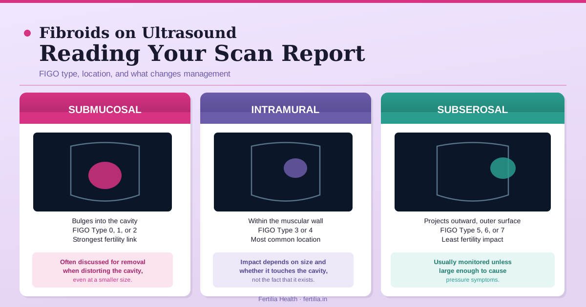
Submucosal fibroids grow inward, bulging into the uterine cavity, the space where a pregnancy would sit. These are the least common type but carry the strongest connection to heavy bleeding and to fertility difficulty, because they distort the surface an embryo would need to implant on.
Intramural fibroids sit within the muscular wall of the uterus. This is the most common location. Whether an intramural fibroid matters clinically depends heavily on its size and on how close it sits to the cavity, not on the fact that it exists.
Subserosal fibroids grow outward, on the outer surface of the uterus, projecting away from the cavity. Because they do not touch the endometrial lining, they are the location least likely to affect periods or fertility, even when they grow fairly large.
A pedunculated fibroid, submucosal or subserosal, is one attached to the uterus by a narrow stalk rather than a broad base. The stalk detail matters for planning removal, since a pedunculated fibroid is often easier to remove with less disruption to the surrounding muscle.
If you are holding a fibroid report and are not sure what your specific findings mean for you, a video consultation is the fastest way to get a specific answer rather than a general one.
Dr. Suganya Venkat is available online, pan-India, for a Rs. 399 video consultation. You can share your scan report directly in the conversation.
The FIGO classification: what the number on your report means
If your report includes a number after “FIGO type,” this is the system radiologists and gynaecologists use to describe exactly where a fibroid sits, on a scale from 0 to 8. It was published as part of the FIGO PALM-COEIN classification for causes of abnormal uterine bleeding (Munro MG et al., International Journal of Gynecology & Obstetrics, 2011; PMID 21345435), and it replaced the older, looser habit of just writing “submucosal” or “intramural” with a number that tells the reader precisely how much of the fibroid sits inside the cavity.
Type 0. Entirely inside the cavity, attached by a stalk (pedunculated submucosal). No part of the fibroid is within the muscular wall.
Type 1. Mostly inside the cavity, with less than half of the fibroid extending into the muscular wall.
Type 2. Mostly within the muscular wall, with more than half of the fibroid extending into the wall, but still touching and distorting the cavity.
Type 3. Entirely within the muscular wall, touching the lining of the cavity (endometrium) but not bulging into the cavity space itself.
Type 4. Entirely within the muscular wall, not touching the cavity lining or the outer surface of the uterus at all.
Type 5. More than half within the muscular wall, extending outward toward the outer surface (subserosal component present).
Type 6. Less than half within the muscular wall, mostly projecting outward toward the outer surface.
Type 7. Entirely subserosal, attached by a stalk (pedunculated subserosal), similar to Type 0 but on the outside.
Type 8. A fibroid in a location not otherwise described by Types 0 to 7. Examples include a cervical fibroid or a fibroid attached to a structure other than the uterine wall.
Why the number matters more than the label. Types 0, 1, and 2 all touch or distort the cavity, which is why these are the types most consistently linked to heavier periods and to reduced implantation rates. Types 3 and 4 sit entirely in the wall without touching the cavity, and behave more like the “intramural, not distorting the cavity” fibroids that are usually watched rather than treated. Types 5, 6, and 7 lean progressively more toward the outer surface, and generally have the least effect on periods and fertility, similar to a purely subserosal fibroid.
A report describing “two fibroids: FIGO type 3 and FIGO type 6” is telling you, in shorthand, that one fibroid touches the cavity lining and one sits mostly on the outside. That distinction usually matters more for planning than the fact that there are two.
What “degeneration” and other descriptive terms mean
Fibroids can outgrow their blood supply, particularly larger ones or those that grow quickly (including during pregnancy). This process is called degeneration, and your report may use one of the following terms.
Hyaline degeneration. The most common type. The fibroid tissue is gradually replaced by a smooth, uniform material. Usually an incidental finding with no specific symptoms, seen often in larger, older fibroids.
Cystic degeneration. Fluid-filled spaces form within the fibroid as tissue breaks down. Appears as dark, fluid-containing areas within the solid fibroid on ultrasound.
Red (carneous) degeneration. Sudden bleeding within the fibroid, most classically seen in pregnancy as a fibroid rapidly outgrows its blood supply under the influence of pregnancy hormones. This type is more likely to cause sudden, localised pain, and is the degeneration type most likely to prompt an urgent review rather than routine follow-up.
Calcific degeneration. Calcium deposits form within older fibroids, often in women closer to or past menopause. Appears as bright, dense areas with acoustic shadowing on ultrasound (similar in appearance to how a gallstone or kidney stone looks on scan). Usually a marker of a longstanding, stable fibroid rather than a new concern.
Degeneration on its own is a description of what the fibroid tissue looks like, not a diagnosis that changes management by itself. Red degeneration causing pain is the exception, and that is usually clear from symptoms at the time, not just the scan.
What size means for management
There is no single centimetre threshold that applies to every fibroid, because location changes what a given size means.
For submucosal (Type 0-2) fibroids, even a relatively small fibroid, sometimes under 2 cm, is often considered for removal if it is visibly distorting the cavity and the woman has heavy bleeding or is trying to conceive, because the cavity distortion is the issue, not the size.
For intramural (Type 3-4) fibroids, most gynaecologists begin discussing removal when a fibroid reaches around 4 to 5 cm and is visibly affecting the cavity or causing bulk symptoms (pressure, a feeling of fullness, bladder frequency). Smaller intramural fibroids not touching the cavity are typically monitored with a repeat scan rather than treated.
For subserosal (Type 5-7) fibroids, size matters less and symptoms matter more. A large subserosal fibroid causing pressure symptoms may be considered for removal regardless of the exact centimetre count, while an asymptomatic subserosal fibroid of the same size is often simply followed.
A report noting the fibroid is “stable in size compared to prior scan” is a reassuring line. Most fibroids grow slowly, if at all, and a stable finding over one to two years of follow-up is a common, unremarkable result.
When a fibroid report changes what happens next
Not every fibroid finding needs a change in plan. As a general pattern based on the location and FIGO type on your report:
Usually monitored, not treated: small intramural fibroids (Type 3-4) not distorting the cavity, small subserosal fibroids (Type 5-7) without symptoms, and any fibroid that has remained stable on repeat scan.
Usually discussed for treatment: submucosal fibroids (Type 0-2) visibly distorting the cavity, particularly alongside heavy periods, difficulty conceiving, or recurrent early pregnancy loss; any fibroid causing significant pressure symptoms regardless of type; and fibroids that have grown notably between scans.
Usually needs prompt review: sudden, severe pain with a known fibroid (possible red degeneration or, rarely, torsion of a pedunculated fibroid), or a rapidly enlarging fibroid, particularly in pregnancy.
The full clinical picture, your symptoms, your fertility plans, and how the fibroid has behaved over time, sits alongside the report to decide what happens next. The fertility-specific version of this discussion, including which fibroid types genuinely reduce conception rates and what the evidence shows about removal, is covered in our companion post on conceiving naturally with fibroids.
For women considering surgical removal, fibroid surgery (myomectomy) cost and recovery in India walks through what hysteroscopic versus laparoscopic or open myomectomy involves at each FIGO type.
If your heavy periods are the reason you were scanned in the first place, our guide on heavy periods after 35 separates out fibroids, adenomyosis, and PCOS as distinct causes, since a report can sometimes show more than one.
Frequently asked questions
What does “FIGO type” mean on a fibroid ultrasound report?
FIGO type is a number from 0 to 8 that describes exactly where a fibroid sits relative to the uterine cavity and wall, using the FIGO PALM-COEIN classification system. Lower numbers (0-2) mean the fibroid touches or bulges into the cavity; middle numbers (3-4) mean it sits entirely within the muscular wall; higher numbers (5-7) mean it leans toward the outer surface of the uterus. Type 8 covers unusual locations such as the cervix.
What is a bulky uterus with fibroids?
“Bulky uterus” describes a uterus larger than the typical size for a woman who has not carried a pregnancy, most often because of the fibroids present. It is a size observation on the report, not a separate condition, and does not by itself indicate a specific treatment is needed.
Which fibroid location affects fertility the most?
Submucosal fibroids (FIGO Type 0, 1, or 2), the ones that touch or bulge into the uterine cavity, have the strongest and most consistent link to reduced implantation rates and early pregnancy loss. Intramural fibroids not touching the cavity, and subserosal fibroids on the outer surface, generally have a much smaller effect on fertility.
Does the number of fibroids matter more than their size?
Not on its own. A uterus with several small fibroids is not automatically more concerning than one with a single larger fibroid. What matters most is whether any individual fibroid, regardless of how many are present alongside it, is distorting the uterine cavity or causing symptoms.
What does fibroid degeneration mean, and is it serious?
Degeneration means the fibroid has outgrown part of its blood supply and the tissue is changing character (hyaline, cystic, calcific, or red degeneration). Most types are incidental findings that do not change management. Red (carneous) degeneration, most often seen in pregnancy, can cause sudden localised pain and is the type most likely to need prompt assessment.
Do all fibroids need to be removed?
No. Most fibroids, particularly small intramural fibroids not touching the cavity and subserosal fibroids without symptoms, are safely monitored with periodic scans rather than treated. Removal is usually discussed when a fibroid distorts the cavity, causes heavy bleeding or pressure symptoms, or is relevant to fertility plans.
How often should a fibroid be rescanned?
There is no single rule for every fibroid; your gynaecologist will set the interval based on the type, size, and your symptoms. A common pattern for a stable, asymptomatic fibroid is a repeat scan at six to twelve months to confirm it has not grown or changed. Fibroids relevant to fertility or causing symptoms are typically followed more closely.
If you have a fibroid report and are unsure what your specific FIGO type and size mean for your periods or fertility plans, speaking with an OB-GYN gives you a specific answer rather than a general one.
Dr. Suganya Venkat is available online, pan-India, for a Rs. 399 video consultation. You can share your scan report directly in the conversation and get an answer specific to your findings.
Dr. Rajashree NS is a Consultant Radiologist with an MD in Radio-diagnosis from Sree Mookambika Institute of Medical Sciences. She interprets pelvic and fertility ultrasound reports as part of the Fertilia Health team.