The message that brings most women to this page started at a scan or a consultation. The ultrasound showed a thickened lining. Or there was bleeding that did not match the usual pattern. Or the doctor said three words that feel much larger than they are: you need a biopsy.
That phrase carries a weight that other medical instructions do not. It implies uncertainty, something possibly serious, something the doctor is not quite saying.
I want to address that directly before anything else.
An endometrial biopsy is a diagnostic test. It tells us what the cells of your uterine lining look like under a microscope. The vast majority of biopsies done in clinical practice, in women referred because of abnormal bleeding or a thickened appearance on scan, come back showing benign tissue. Most results show normal endometrium or a condition like hyperplasia that is managed without surgery.
The biopsy is not a treatment. It is information. And the information it gives us is, most of the time, reassuring.
This guide covers why the test is recommended, what the Pipelle procedure involves, what it actually feels like, what it costs in India, and how to interpret whatever result comes back.
Why Is an Endometrial Biopsy Recommended?
A biopsy is recommended when we need to look at the actual cells of the uterine lining, not just its appearance or thickness on a scan. There are four clinical situations where this becomes necessary.
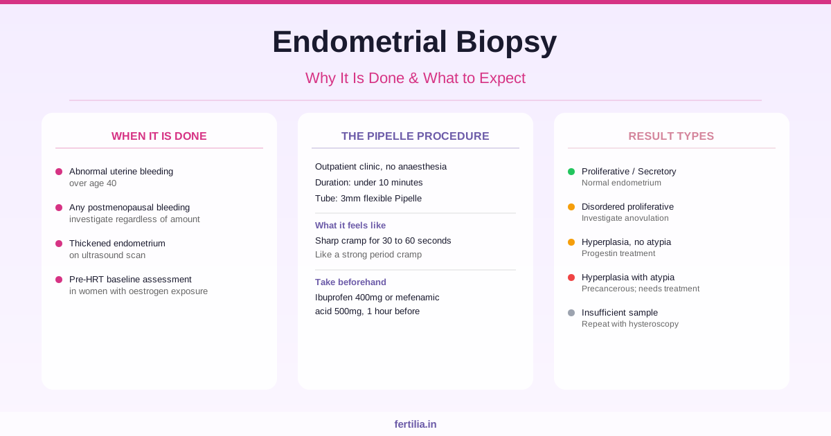
Abnormal uterine bleeding (AUB) in women over 40. Irregular, heavy, or intermenstrual bleeding that does not settle with initial investigation in a woman over 40 warrants endometrial sampling. This is not because we suspect cancer in every case. It is because the risk of endometrial hyperplasia and carcinoma increases with age, and the only way to rule it out with confidence is to examine the tissue directly. Women under 40 with persistent AUB may also be sampled if they have had prolonged oestrogen exposure from PCOS-related anovulation, obesity, or certain medications, or if they have gone many months without a natural period.
Postmenopausal bleeding. Any vaginal bleeding that occurs one year or more after the last menstrual period requires investigation. The clinical rule is firm: postmenopausal bleeding is investigated as a possible endometrial problem until a cause is confirmed. In practice, the large majority of women investigated for postmenopausal bleeding do not have cancer. A thin endometrium (less than 4mm on transvaginal ultrasound) with reassuring clinical findings often provides sufficient information; a thicker or irregular lining requires sampling.
Thickened or irregular endometrium on ultrasound. The scan shows the lining’s thickness and contour but not what the cells are doing. An endometrial thickness above the threshold for your hormonal status, or an irregular, heterogeneous appearance, prompts a biopsy to understand what is underneath. For more on what scan measurements mean in context, see our guide to Endometrial Thickness: Trilaminar Pattern and Normal Ranges.
Pre-HRT baseline assessment. In certain clinical situations, particularly when a woman has had prolonged unopposed oestrogen exposure, a biopsy is done before starting hormone replacement therapy to confirm that the lining is normal at the outset.
Pipelle Biopsy, Hysteroscopy-Guided Biopsy, or D&C: Which One?
There are three ways to obtain an endometrial sample, and the choice depends on the clinical situation.
The Pipelle biopsy (office biopsy) is the standard starting point and what most women will actually have. A Pipelle is a thin, flexible plastic tube, roughly 3mm in diameter, passed through the cervix into the uterine cavity. A small piston inside creates suction, and the tube is gently rotated and moved to collect a tissue sample. The sampling takes under two minutes once the tube is in position, and the whole appointment is outpatient, with no anaesthesia required.
Hysteroscopy-guided biopsy is used when the Pipelle does not yield an adequate sample, which happens in approximately 10 to 15% of cases and is more common in postmenopausal women with a narrowed cervix. It is also used when the ultrasound shows a focal abnormality (a polyp, a small fibroid, or a localised thickening) that needs to be directly visualised and targeted rather than randomly sampled. This requires a small camera passed into the uterine cavity under local or general anaesthesia, and is done in a procedure room or theatre. For cost information and what to expect, see our Hysteroscopy Cost in India 2026 guide.
Dilatation and curettage (D&C) is now rarely used purely for diagnosis. It is usually reserved for situations where treatment and sampling happen together, such as when a uterine polyp is being removed at the same time as the lining is sampled.
For most women referred for an endometrial biopsy, a Pipelle in an outpatient clinic is what will happen. It is quicker, done without general anaesthesia, and has a sensitivity of over 90% for detecting endometrial hyperplasia and carcinoma when the sample is adequate.
What the Pipelle Procedure Feels Like
This is the question I hear most often from women preparing for the test.
The Pipelle biopsy causes discomfort. For most women, it is a sharp cramping sensation, similar to a strong period cramp, lasting 30 to 60 seconds as the sample is collected. Some women feel a brief, stronger cramping as the tube passes through the cervix. It is uncomfortable, but it is short.
Women who have not had a vaginal delivery and postmenopausal women with a narrowed cervical opening tend to find the procedure more uncomfortable than women who have delivered vaginally. If you fall into either group, tell your doctor beforehand so that appropriate adjustments can be made.
Taking a painkiller before the appointment helps. A standard dose of ibuprofen (400mg) or mefenamic acid (500mg) taken 45 to 60 minutes before the procedure reduces cramping during the sampling. Discuss this with your doctor first, particularly if you have kidney problems, asthma, or are taking blood thinners or other regular medications.
During the procedure: You lie in the same position as a routine gynaecological examination. The doctor places a speculum, cleans the cervix, and then passes the Pipelle. Being asked to cough or breathe slowly as the tube enters the cervix can reduce discomfort briefly. The sampling itself takes about 30 to 60 seconds.
Afterward: Mild to moderate cramping often continues for a few hours, similar to the first day of a period. Light spotting or a brownish discharge for one to three days is normal. Most women return to normal activity the same day. Avoid sexual intercourse and swimming for 48 hours after the procedure. Contact your doctor if you develop fever, heavy bleeding, or significant pelvic pain in the days that follow.
If you have specific questions about what to expect before your procedure, Dr. Suganya Venkat, OB-GYN with over 15 years of clinical experience, is available for a video consultation for women across India.
Endometrial Biopsy Cost in India (2026)
The cost varies significantly by setting, and it is worth understanding what each quote typically includes or excludes.
| Setting | Approximate cost |
|---|---|
| Government hospital or AIIMS | Rs. 250 to 1,500 (procedure); Rs. 500 to 3,000 including pathology |
| CGHS-empanelled hospital (CGHS beneficiaries) | Rs. 2,040 to 3,000 depending on city tier |
| Private clinic or mid-range hospital (Pipelle, outpatient) | Rs. 3,000 to 10,000 |
| Corporate hospital chain (package including consultation, materials, and follow-up) | Rs. 18,000 to 80,000 |
Prices verified August 2026. Always confirm costs directly with your hospital or clinic at the time of booking.
The histopathology fee (the lab processing fee for the tissue sample) is often billed separately. This typically adds Rs. 800 to 2,500. Ask whether the pathology report is included in the quote before you book.
If the biopsy needs to be done under hysteroscopy guidance because the Pipelle was insufficient or a focal lesion was found, the cost increases considerably. Hysteroscopy-guided biopsy in private settings typically falls in the range of Rs. 15,000 to 50,000.
Health insurance may cover the diagnostic biopsy procedure depending on your policy. Check with your insurer before the appointment.
How to Read Your Biopsy Results
Histopathology results typically take 3 to 7 working days from private labs and 5 to 10 working days from government facilities. The report describes what the pathologist found in the tissue sample. Here is what the commonly seen terms mean.
Proliferative endometrium. The lining is in its growth phase, stimulated by oestrogen. This is normal in the first half of the menstrual cycle. A benign finding.
Secretory endometrium. The lining has responded to progesterone and is in the second half of the cycle. Also normal and benign.
Disordered proliferative endometrium. The lining cells are proliferating but not in a fully organised pattern. This is typical of anovulatory cycles, where ovulation does not occur and the lining stays under sustained oestrogen stimulation without a progesterone phase. It is not cancerous. It is a signal that the cycle needs attention, whether from PCOS-related anovulation, perimenopause, or other hormonal factors.
Endometrial hyperplasia without atypia. The lining has overgrown because of excess oestrogen without adequate progesterone to counterbalance it. Under the microscope, the cells still look structurally normal. This is common in women with anovulatory PCOS, women who are overweight, and perimenopausal women. It responds well to progestin treatment. A Mirena intrauterine system or oral progestins are used, with a repeat biopsy at 3 to 6 months to confirm the lining has normalised. With appropriate treatment, the risk of this progressing to cancer is very low. For context on Mirena as a treatment approach, see Mirena: The Hysterectomy Alternative for Heavy Periods.
Endometrial hyperplasia with atypia (also called atypical hyperplasia or EIN, endometrial intraepithelial neoplasia). The cells show structural abnormalities under the microscope. This is classified as a precancerous condition. It requires treatment: either high-dose progestins for women who wish to preserve fertility, or hysterectomy for women who do not. This is not cancer, but it needs prompt specialist management.
Endometrial carcinoma. Cancer of the uterine lining. The report will describe the grade and type, most commonly endometrioid. This leads to referral to a gynaecological oncologist for staging and a treatment plan. Early-stage endometrial carcinoma, which is what is typically found when it is caught through investigation of bleeding symptoms, has a good prognosis when treated promptly. For more on the risk picture: Endometrial Cancer: Symptoms, Risk Factors and Warning Signs.
Insufficient sample. No interpretable tissue was obtained. This happens in 10 to 15% of Pipelle biopsies, most commonly in postmenopausal women or those with a narrowed cervix. It does not mean something is wrong with the lining. It means the procedure needs to be repeated with a different technique, usually hysteroscopy.
If You Have Heavy or Irregular Periods After 35
If your periods have changed over the past one to three years (heavier, more irregular, or with bleeding between cycles), an endometrial biopsy is one part of a broader investigation. The biopsy rules out hyperplasia and carcinoma; the ultrasound identifies structural causes like fibroids, adenomyosis, or polyps; and blood tests assess thyroid function, clotting, and hormonal status.
The conditions most commonly responsible for AUB at this stage of life are benign and treatable. For more on the full picture: Heavy Periods After 35: Fibroids, Adenomyosis or PCOS? and Heavy Periods: Causes and Every Treatment Option.
For a broader resource on period health and when changes are worth investigating, the Period Health guide at Fertilia covers what to track and what to bring to your next appointment.
If your biopsy result has come back and you are unsure what it means or what to do next, a video consultation covers exactly that: what the result means for you specifically, what the options are, and what the next step should be.
WhatsApp Dr. Suganya Venkat to discuss your biopsy result
Frequently Asked Questions
Is an endometrial biopsy painful?
Yes, briefly. The Pipelle causes a sharp cramping sensation, similar to a strong period cramp, lasting 30 to 60 seconds as the tissue is collected. Most women describe it as uncomfortable but manageable, particularly with ibuprofen or mefenamic acid taken an hour before. Postmenopausal women and women who have not delivered vaginally tend to find it more uncomfortable than others. Let your doctor know in advance so they can plan accordingly.
Can I go to work after an endometrial biopsy?
Most women return to normal activity the same day. Light cramping and spotting for a few hours is common. If your work involves physical exertion, or you know period pain typically affects you significantly, having the afternoon free gives you flexibility. There are no formal restrictions on returning to desk work.
How long does the whole appointment take?
The appointment takes around 15 to 30 minutes in total, including the consultation, positioning, and procedure. The sampling itself takes under two minutes once the Pipelle is in position.
My scan shows thickened endometrium. Does that mean cancer?
Not at all. A thickened endometrium on scan is a finding that needs investigation, not a diagnosis. Many causes of endometrial thickening are entirely benign: normal cycle variation, PCOS-related anovulation, polyps, fibroids, or hormonal fluctuations around perimenopause. The biopsy tells us what the cells are actually doing, which the scan alone cannot show.
Is endometrial biopsy dard hota hai? (Is the biopsy painful in Hindi?)
Yes, briefly. It causes discomfort similar to strong period cramps for about a minute during the sampling. Taking a painkiller (ibuprofen or mefenamic acid) an hour before the procedure helps considerably. After the procedure, light cramping and spotting for a few hours is normal.
My biopsy showed endometrial hyperplasia without atypia. Should I be worried?
Hyperplasia without atypia is a benign, non-cancerous overgrowth of the uterine lining. It happens when the lining has been under sustained oestrogen stimulation without enough progesterone to balance it. This is common with PCOS-related anovulation, obesity, and perimenopause. It responds well to progestin treatment and, with follow-up, does not progress to cancer in the vast majority of cases.
What if the biopsy comes back as insufficient or no tissue found?
An inadequate sample means the tube could not collect enough tissue for analysis. This happens in 10 to 15% of Pipelle biopsies and is more common in postmenopausal women. If the clinical concern is significant, the investigation continues with a hysteroscopy, which directly visualises the cavity and takes a targeted sample. An insufficient result is not a bad sign; it is a technical outcome that directs the next step.