You have come from the anatomy scan or a routine check at twenty weeks. The report is in your hand and somewhere in the findings section it reads: “Cervical length 22 mm. Short cervix noted. Please correlate clinically.” You were not expecting this. Your scan appointment was about checking your baby’s heart chambers and spine; no one prepared you for a note about your cervix. Now you are searching for what 22 mm means.
I am a radiologist. I report these measurements every week. The number on your report is important enough to be taken seriously, and it is calm enough that panic is not the right response to it either. Let me explain what that measurement is, what the thresholds mean, and what happens next, so you can have a useful conversation with your obstetrician instead of spending the next several days at the wrong end of a search engine.
What this guide covers:
- What cervical length is and why it is measured on ultrasound
- Why the transvaginal scan (TVS) gives the most accurate reading
- Normal cervical length ranges by trimester
- What short cervix means and at what threshold the term applies
- Terms that appear on the report: funnelling, beaking, sludge
- What the monitoring and management pathway looks like
- How this finding is handled in India
What Cervical Length Is and Why It Matters
The cervix is the lower, narrow portion of the uterus. For most of pregnancy it stays firm, closed, and long, acting as a plug that holds the growing baby safely inside. It only shortens and opens when labour is approaching at term.
Cervical length is the physical measurement of that plug from the internal os (the opening between the uterus and the cervical canal) to the external os (the opening at the lower end of the cervix, at the top of the vagina). In a healthy mid-pregnancy uterus, this length is typically between 30 and 45 mm.
The reason radiologists and obstetricians measure it is straightforward: a shorter cervix in the second trimester is one of the strongest ultrasound predictors of spontaneous preterm birth. The landmark study by Iams and colleagues, published in the New England Journal of Medicine in 1996 (PMID 8569824), measured cervical length in over 2,900 women at 24 weeks. Women with a cervical length below 25 mm had a significantly higher risk of delivering before 35 weeks than those with a longer cervix. The shorter the cervix, the greater the risk, and the risk increases continuously rather than switching on at a single cut-off point.
This means the measurement has real predictive value, and it is why your report has flagged it. A measurement of 22 mm at twenty weeks is not a normal finding. But it is also not a sentence. It is a prompt to act, to monitor, and to consider evidence-based interventions that work.
Why the Transvaginal Scan Is the Standard for Cervical Length
Your routine anatomy scan may have been done abdominally, with the probe placed on your belly. Abdominal ultrasound gives good views of the baby but is not reliable for measuring the cervix accurately. The bladder’s fullness or emptiness changes how the cervix appears. The angle is less direct. The measurement can appear falsely reassuring.
Cervical length is measured by transvaginal ultrasound (TVS): the probe is gently placed in the vagina, a few centimetres from the cervix, so the view is direct and the measurement is not affected by the abdominal wall, the bladder, or the baby’s position. The procedure is safe in pregnancy. It does not stimulate labour or cause any harm to the cervix.
If your routine anatomy scan was done abdominally and the sonographer had difficulty seeing the cervix clearly, or the measurement was borderline, your obstetrician will almost certainly ask for a dedicated transvaginal assessment. This is standard, not alarming, and it produces a measurement you can rely on.
On a transvaginal scan, the cervix appears as a linear structure. The radiologist identifies the internal and external os, places callipers at each end, and records the straight-line distance between them. The average of three measurements, taken in sequence, is reported. The standard position is with the patient’s bladder empty, with no pressure on the cervix from the probe.
Normal Cervical Length by Trimester
Cervical length naturally shortens as pregnancy progresses, particularly in the third trimester as the body begins preparing for labour. The ranges below are the values used in clinical practice across published obstetric literature.
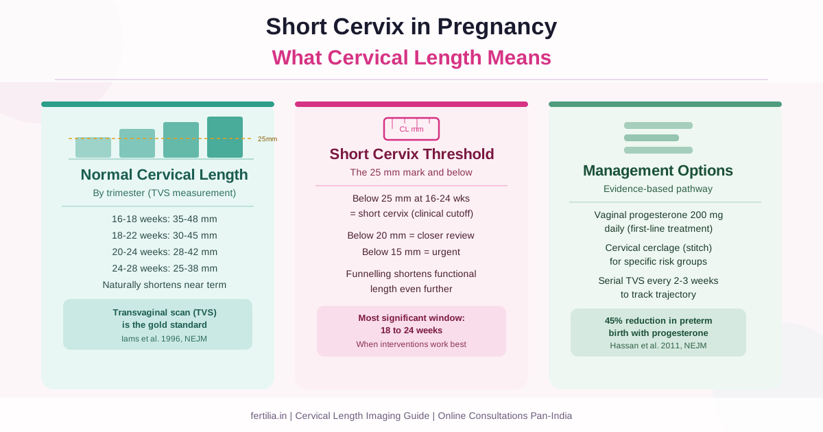
| Period | Typical Range | Threshold of Concern |
|---|---|---|
| 16 to 18 weeks | 35 to 48 mm | Below 25 mm |
| 18 to 22 weeks | 30 to 45 mm | Below 25 mm |
| 20 to 24 weeks | 28 to 42 mm | Below 25 mm |
| 24 to 28 weeks | 25 to 38 mm | Below 20 mm |
| 28 to 32 weeks | 20 to 35 mm | Below 15 mm |
The most clinically significant window is 18 to 24 weeks. This is when:
- Spontaneous preterm birth risk is most strongly predicted by cervical length
- Interventions such as progesterone and cerclage have the best evidence for benefit
- Funnelling and other structural changes on the report are most meaningful
A measurement of 35 mm at 22 weeks is reassuring. A measurement of 22 mm at the same point is short enough to warrant surveillance and possibly treatment. A measurement of 15 mm or below at 24 weeks is short enough that your obstetrician will likely want to discuss cerclage alongside other measures.
What “Short Cervix” Means
The clinical definition of short cervix, based on consensus from major international obstetric bodies, is a cervical length below 25 mm measured between 16 and 24 weeks of pregnancy by transvaginal ultrasound.
This threshold comes from the data. The Iams 1996 study and subsequent large cohort studies consistently show that the 25 mm mark separates the population of women at meaningfully elevated preterm risk from those with lower risk. It is not a sharp cliff, it is a point on a continuum, but it is the point at which monitoring and treatment conversations begin.
Some centres use 20 mm as the threshold for more urgent intervention, particularly for cerclage decisions. You may see different thresholds used in different hospitals. The important thing is to understand that 25 mm is the benchmark, and measurements closer to 15 mm or below represent a more urgent category.
If your report reads 22 mm, you are below the 25 mm threshold. If it reads 28 mm, you are above it but close enough that a repeat scan in two to three weeks is reasonable. If it reads 18 mm, your obstetrician will likely want to move quickly.
Terms That May Appear on Your Cervical Length Report
Funnelling
Funnelling refers to the internal os (the upper opening of the cervix) beginning to open, with amniotic fluid descending into the upper part of the cervical canal. On ultrasound this creates a V-shaped or U-shaped appearance at the top of the cervix.
The effective cervical length reported is measured from the bottom of the funnel (where the cervical canal closes) to the external os, not the total length from the original internal os. Funnelling effectively shortens the functional cervix even when the overall anatomical length appears reasonable.
A small amount of funnelling is sometimes seen at term as labour approaches, but funnelling detected before 24 weeks, particularly with a short residual cervical length, is a marker of cervical incompetence and raises preterm birth risk. If your report mentions funnelling, this is a finding that warrants urgent discussion with your obstetrician.
Beaking
Beaking is a variant of funnelling where the top of the cervical canal takes on a triangular, beak-like shape on ultrasound. It is sometimes considered a less advanced stage than a fully formed funnel but is reported for the same reason: it represents early effacement of the internal os.
Sludge
Amniotic sludge is a hyper-reflective (bright) particulate material seen in the fluid within the lower uterine segment or the funnel, visible on ultrasound. It is thought to represent an aggregation of inflammatory material and has been associated with intra-amniotic infection and an elevated risk of preterm birth when detected alongside a short cervix.
A report that mentions sludge in the context of a short cervix is an urgent finding. Your obstetrician will likely want to assess you the same day or within 24 hours.
What Happens After a Short Cervix Finding
The management of a short cervix depends on the measurement, the stage of pregnancy, your obstetric history, and whether additional findings such as funnelling are present. I can describe the three main options your obstetrician may discuss with you.
Vaginal Progesterone
For women with a short cervix detected on mid-trimester ultrasound and no prior history of preterm birth, vaginal progesterone (a micronised progesterone pessary or gel, typically 200 mg daily) is the first-line evidence-based treatment.
Two large randomised controlled trials have established this approach. Fonseca and colleagues (2007, PMID 17671254, published in the New England Journal of Medicine) showed that 200 mg daily vaginal progesterone reduced the rate of preterm birth before 34 weeks in women with a cervical length below 15 mm. Hassan and colleagues (2011, PMID 21472815, the PREGNANT trial, published in Ultrasound in Obstetrics & Gynecology) confirmed this using vaginal progesterone gel in women with a cervical length of 10 to 20 mm: preterm birth before 33 weeks fell from 16.1 percent to 8.9 percent, a 45 percent relative reduction. The FIGO working group and the Society for Maternal-Fetal Medicine both recommend offering vaginal progesterone to women with a singleton pregnancy and a cervical length below 25 mm at 24 weeks.
In India, micronised progesterone is prescribed as a vaginal pessary (common brand names include Utrogestan and Gestofit). Note that Duphaston, which is often mentioned in the same conversation, is dydrogesterone, a different molecule taken orally, and it is not what these short-cervix trials tested. It is taken nightly, is generally well tolerated, and continues until 36 to 37 weeks or until the obstetrician advises otherwise.
Vaginal progesterone does not cure a short cervix. It does not make the cervix physically longer. It reduces the risk of preterm birth through a mechanism involving uterine quiescence, and it is the most accessible and least invasive starting point for most women.
Cervical Cerclage (Cervical Stitch)
Cerclage is a stitch placed around the cervix under anaesthesia to provide mechanical support. It is not offered to every woman with a short cervix. The two scenarios in which it is most consistently recommended are:
History-indicated cerclage: Women with three or more second-trimester losses or preterm births before 34 weeks, thought to be due to cervical insufficiency. In this group, cerclage is placed prophylactically at 12 to 14 weeks regardless of cervical length.
Ultrasound-indicated cerclage: Women with a singleton pregnancy, no prior preterm birth, and a cervical length below 25 mm detected on TVS between 16 and 24 weeks. The evidence for cerclage in this group is less uniformly positive than for progesterone, and many obstetricians now use progesterone first and consider cerclage if the cervix continues to shorten. Women with a prior preterm birth AND a short cervix on this scan have the strongest evidence for cerclage benefit.
The decision is your obstetrician’s to make after reviewing your full history. If cerclage is being considered, you can also read the dedicated guide on what a cervical stitch involves and what to expect, which covers the procedure, recovery, and delivery in more detail.
Serial Cervical Length Monitoring
If your cervical length is between 25 and 30 mm, or if it is your first short cervix finding and your obstetrician wants to confirm the measurement before committing to treatment, the next step is a repeat transvaginal scan in two to three weeks.
A cervical length that is stable at 25 to 30 mm with no funnelling over two serial scans in a woman without other risk factors may be monitored without intervention. A cervical length that decreases from scan to scan is a more dynamic finding and will usually prompt treatment.
Serial monitoring allows your obstetrician to track the trajectory, not just a single snapshot. A cervix at 28 mm that stays at 28 mm is different from a cervix at 28 mm that is 22 mm three weeks later.
My cervical length report has me worried. Can I speak with someone? Write to us on WhatsApp.
Cervical Length in India: Practical Points
Where is the scan done?
A transvaginal cervical length scan is available at most private obstetric ultrasound centres and at larger government hospitals. If your anatomy scan was done at a general radiology centre, ask for the cervical length to be checked by TVS specifically: not all centres routinely measure it abdominally, and an abdominal measurement alone is not reliable enough to act on.
What does it cost?
A dedicated TVS cervical length assessment, booked separately from a routine scan, typically costs between Rs. 800 and Rs. 1,500 at private centres in major cities. At government hospitals and some teaching hospital antenatal clinics, it is available within the regular antenatal scan package at no additional cost.
Vernacular terminology
In Hindi, the cervix is sometimes referred to as the gardan (neck) of the uterus, or simply as the cervix. A short cervix may be described as choti gardan in informal conversation. The phrase gardan ki lambai is the literal translation of cervical length. If you are attending a government hospital where reports are summarised verbally in Hindi or Tamil, asking about the gardan ki lambai in Hindi (or keel-kandam neeam in Tamil, Roman transliteration) will help you identify the measurement on your report.
The preterm labour connection
A short cervix is one of the factors that raises the risk of preterm labour. If you have a confirmed short cervix, your obstetrician should discuss with you which warning signs to watch for and when to present to the hospital, regardless of whether progesterone or cerclage has been started.
How This Reading Connects to Other Scans
If you are trying to understand your full antenatal scan picture, the following guides may also be useful:
- Anomaly scan (TIFFA) at 20 weeks: the same scan at which cervical length is often first checked
- Transvaginal scan in pregnancy: what a TVS involves and when it is used
- Growth scan and Doppler: reading the third-trimester report: the later pregnancy monitoring scan
- High-risk pregnancy care: how short cervix fits into the broader high-risk antenatal pathway
FAQ
What is a normal cervical length at 20 weeks?
A normal cervical length at 20 weeks on transvaginal ultrasound is approximately 35 to 40 mm, with most guidelines using 25 mm as the lower threshold below which a pregnancy is considered at increased risk of preterm birth. A length above 25 mm at this point is reassuring. A length below 25 mm warrants discussion with your obstetrician about monitoring and treatment.
I have a cervical length of 22 mm at 22 weeks. Is this serious?
22 mm at 22 weeks is below the 25 mm threshold that marks a short cervix and will prompt clinical action. It is serious enough to take seriously: your obstetrician will likely discuss vaginal progesterone and repeat scanning, and may also discuss cerclage depending on your history. It is not so short that preterm delivery is inevitable; women with this measurement who receive appropriate management carry pregnancies to term regularly.
Can I have a normal delivery if I have a short cervix?
Yes. A short cervix, once managed with progesterone, cerclage, or close monitoring, does not by itself prevent a vaginal delivery. What matters is that the cervix stays closed until the baby is ready, and that any shortening trend is identified and managed early. Most women with a short cervix who receive appropriate treatment have healthy pregnancies.
What is the difference between cervical length and cervical incompetence?
Cervical incompetence (also called cervical insufficiency) is a clinical diagnosis made when a woman has experienced one or more painless second-trimester pregnancy losses or preterm births without contractions, thought to be because her cervix opened silently. A short cervix on ultrasound is one finding that can support this diagnosis, but the clinical history matters too. A short cervix discovered on a routine mid-trimester scan in a first pregnancy is not automatically the same as a diagnosis of cervical incompetence.
Will I need the cervical length checked in my next pregnancy?
If you have a short cervix in this pregnancy, yes. A history of a short cervix or preterm birth significantly increases your risk in subsequent pregnancies, and early TVS surveillance from 16 weeks is standard in women with this history. Some obstetricians will also offer history-indicated cerclage in the next pregnancy before any scan shows shortening, if the clinical picture suggests it.
Is vaginal progesterone the same as the progesterone given to prevent miscarriage?
Progesterone is prescribed at several different points and for different reasons in pregnancy. The vaginal progesterone given for a short cervix (typically 200 mg daily from the diagnosis until 36 to 37 weeks) is a higher and longer course than the progesterone sometimes prescribed in the first trimester for threatened miscarriage or luteal phase support. They are the same molecule but used in different clinical contexts. Always follow your obstetrician’s specific prescription, which will match your situation.
Gardan ki lambai pregnancy mein normal kya hoti hai? (What is normal cervical length in pregnancy?)
20 week ki pregnancy mein normal gardan ki lambai (cervical length) approximately 35 to 40 mm hoti hai. 25 mm se neeche ki lambai ek short cervix mani jati hai, aur iske liye doctor se milna zaroori hai. Yeh lambai transvaginal scan (TVS) se sabse sahi tarike se mapi jati hai.
If your scan report mentions a short cervix or a cervical length below 25 mm, talk to us. Write to us on WhatsApp and we will help you understand the next steps.
Dr. Rajashree NS is a Consultant Radiologist with an MD in Radio-diagnosis. She brings a radiologist’s perspective to reading obstetric and gynaecological scan reports, translating the numbers and terms into plain language for the patient holding the printout.