A woman comes home carrying her anomaly scan report. The radiologist said very little during the examination. The printout reads: “TIFFA scan done. No fetal anomaly detected. Normal study.” She is relieved, but uncertain. What exactly was checked? What does “normal study” cover, and what would the report have said if something looked different?
I am a radiologist. I perform TIFFA scans daily and write reports in this language. This guide translates a standard TIFFA report into plain words, walks through what the scan examines system by system, and explains what soft markers mean when one appears on an otherwise normal scan.
What this guide covers:
- What the TIFFA is, and how it differs from the Level 1 scan
- Why it is done at 18 to 22 weeks
- The fetal systems the scan checks, one by one
- Fetal biometry and what your measurements mean
- What “No fetal anomaly detected” and “Normal study” mean in practice
- Soft markers: what they are, how common they are, and why context matters
- When a follow-up scan is recommended
What Is the TIFFA Scan?
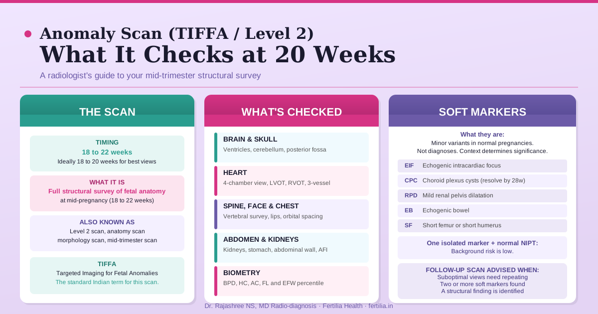
TIFFA stands for Targeted Imaging for Fetal Anomalies. It is the name used in India for the mid-trimester structural survey. Other countries call it the anatomy scan, level 2 scan, morphology scan, anomaly scan, or mid-trimester scan. The names differ; the examination is the same.
The TIFFA is a standard part of antenatal care. It is done between 18 and 22 weeks of pregnancy, with 18 to 20 weeks considered the best window for most views. By 18 weeks, fetal organs have developed enough structure to be assessed clearly on ultrasound. After 22 weeks, certain structures become harder to image as the baby grows larger and lies in more fixed positions.
The TIFFA is not a chromosome test. It does not replace NIPT, the double marker test, or amniocentesis. Those tests assess chromosomal risk through blood or amniotic fluid samples. The TIFFA assesses fetal structure through imaging. Both have a role in antenatal surveillance, and they answer different questions.
For women who had a transvaginal scan or a dating scan in the first trimester, that earlier scan assessed viability, gestational age, and the nuchal translucency measurement. The TVS and its place in early pregnancy imaging covers what is visible before the TIFFA window. The TIFFA goes further, examining each organ system in detail.
Why 18 to 22 Weeks?
The timing reflects what ultrasound can reliably detect at different stages of pregnancy.
Before 18 weeks, some cardiac structures, particularly the ventricular outflow tracts, may not be clearly visualised. The posterior fossa (the space behind the cerebellum) is also better assessed after 18 weeks. Neural tube defects, which affect the spine and brain simultaneously, are most reliably detected before 22 weeks.
After 22 weeks, a structural survey remains useful and can identify most major findings. Some views become progressively harder to obtain as the baby grows. If a TIFFA is missed for any reason, a scan at 24 to 26 weeks can still provide a substantial proportion of the same information, and the radiologist will note any views that were limited.
For more on this, read our guide on Early Pregnancy Scan. The ISUOG Practice Guidelines (International Society of Ultrasound in Obstetrics and Gynecology, updated 2020) recommend the mid-trimester structural survey between 18 and 22 weeks as the standard window for a complete fetal anatomy assessment.
What the TIFFA Checks: Fetal Systems
The scan works through fetal anatomy in a structured sequence. Each system has specific views the radiologist must obtain and document. Here is what is assessed in every standard TIFFA.
Brain and skull
The scan measures the biparietal diameter and head circumference, and checks the lateral ventricles, the cerebellum, the posterior fossa (cisterna magna), and the cavum septum pellucidum (CSP). The CSP is a thin midline structure whose absence can indicate a midline brain defect. The choroid plexus, which produces cerebrospinal fluid, is also assessed.
Face
The fetal profile is checked for the nasal bone. The lips and palate are assessed for cleft lip, which is visible on ultrasound in most cases. Orbital spacing is noted. An isolated cleft palate without cleft lip is harder to detect on ultrasound and is more commonly identified at birth.
Heart
Cardiac anomalies are among the most common structural findings. The TIFFA obtains the four-chamber view, confirming that both atria and both ventricles are present, and checks the left and right ventricular outflow tracts (LVOT and RVOT) to assess for major vessel abnormalities. The three-vessel and three-vessel-trachea view documents the pulmonary artery, aorta, and superior vena cava in relation to the trachea. Fetal heart rate and rhythm are recorded.
Spine
The vertebral column is surveyed from cervical to sacral to look for neural tube defects. Spina bifida produces characteristic secondary brain changes, including the lemon sign (indentation of the frontal bones) and the banana sign (displacement of the cerebellum toward the brainstem). These brain findings often alert the radiologist to a spinal defect before the spine itself is fully assessed.
Lungs and chest
Both lung fields are checked to confirm they are present, symmetric, and appropriately sized relative to the chest cavity. The cardiothoracic ratio and cardiac axis are also noted.
Abdomen and umbilical cord
The abdomen is assessed for the stomach bubble, a dark, fluid-filled structure visible when the baby is swallowing normally. The anterior abdominal wall is examined to exclude defects such as gastroschisis (bowel herniated through a defect beside the cord insertion) or omphalocele (organs covered by membrane but outside the abdomen). The umbilical cord insertion is checked.
The cord is assessed for its vessel composition. A normal cord has two arteries and one vein. A single umbilical artery (SUA), present in roughly 1% of pregnancies, is noted because in a minority of cases it is associated with other structural or chromosomal findings, though in isolation it is often a normal variant.
Kidneys
Both kidneys are identified. The renal pelvis (the collecting structure within each kidney) is measured. A renal pelvis measurement of 4 mm or more at 18 to 22 weeks is classified as mild pyelectasis, one of the most commonly noted findings on the TIFFA. Mild pyelectasis is discussed in the soft markers section below.
Limbs
The femur length is measured as part of biometry. The long bones of all four limbs are assessed for symmetry and appropriate length. Hands and feet are examined where the baby’s position allows. The scan looks for structural findings such as unusual foot positioning or hand abnormalities.
Placenta
The position, appearance, and relationship of the placenta to the internal cervical os are documented. A placenta that overlies the internal os at 20 weeks is flagged for a repeat assessment at 28 to 32 weeks, as the majority of low-lying placentas migrate upward as the uterus grows.
Amniotic fluid
The amniotic fluid index (AFI) or deepest vertical pocket (DVP) is measured. Both very low values (oligohydramnios) and very high values (polyhydramnios) are noted, though borderline values at 20 weeks are common and re-assessed at a later scan when clinically relevant.
For more on this, read our guide on Growth Scan & Doppler in Pregnancy.
Fetal Biometry: The Measurements on Your Report
Every TIFFA report includes fetal biometry: four measurements used to estimate gestational age and fetal size.
- BPD (Biparietal Diameter): the width of the fetal skull at its widest point
- HC (Head Circumference): the outer circumference of the skull
- AC (Abdominal Circumference): the circumference of the abdomen at the level of the liver and stomach
- FL (Femur Length): the length of the thigh bone
From these four values, the estimated fetal weight (EFW) is calculated using the Hadlock formula (Hadlock FP et al., Am J Obstet Gynecol. 1985;151(3):333-337). The EFW and each individual measurement are plotted against gestational age to give a percentile.
A measurement between the 10th and 90th percentile is considered appropriate for gestational age. An EFW or abdominal circumference persistently below the 10th percentile prompts closer monitoring for fetal growth restriction. For more on what a growth-restriction finding means in the third trimester, see our guide on fetal growth restriction and IUGR.
Questions about your TIFFA scan report? Speak with Dr. Rajashree via our online consultation. →
What “Normal Study” Means on Your Report
When your report states “No fetal anomaly detected” or “Normal study,” it means that no structural abnormality was identified in the systems and organs examined, within the resolution limits of ultrasound at 18 to 22 weeks.
There are three qualifications that every radiologist holds in mind when writing this:
Ultrasound has resolution limits. Structures below a few millimetres are not detectable on a standard scan. Small muscular ventricular septal defects (holes in the muscular wall between the two ventricles) are a common example: they may be present but invisible at this gestational age, and many close spontaneously before or after birth. Mild narrowing of the aorta may also be difficult to detect.
Fetal position affects image quality. When the baby is lying in a position that makes certain views difficult to obtain (the face, the spine, or the cardiac outflow tracts are particularly position-dependent), the radiologist will note this in the report and may advise a repeat scan for those specific views.
Some conditions become apparent only in the third trimester. Certain findings, such as late-onset ventriculomegaly (enlargement of the brain’s fluid spaces) or some skeletal dysplasias, are not detectable at 20 weeks. The TIFFA is a thorough assessment at mid-pregnancy, not a guarantee for the full pregnancy.
These qualifications are the reason antenatal care continues with growth scans and monitoring in the third trimester. A normal TIFFA provides strong structural reassurance, which is what it is designed to deliver.
Soft Markers: What They Are and What They Mean
A soft marker is a minor finding on an otherwise normal anomaly scan that can appear in normal pregnancies. Soft markers are not structural abnormalities in themselves. They are called “soft” because they are not independently diagnostic of any condition. Their significance depends entirely on the clinical context in which they are found.
Echogenic intracardiac focus (EIF) A small bright spot, typically in the left ventricle, that appears as a white dot on the scan. It is seen in roughly 3 to 5% of normal fetuses and is thought to represent a minor calcification in the papillary muscle. In a low-risk pregnancy with a normal NIPT result or normal first-trimester combined screening, an isolated EIF is not an indication for further invasive testing (Bromley B et al., J Ultrasound Med. 2002;21(10):1087-1096).
Choroid plexus cysts (CPC) Small cysts within the choroid plexus, seen in roughly 1 to 3% of second-trimester scans. They almost always resolve by 26 to 28 weeks. Their presence in isolation, after normal first-trimester screening or a normal NIPT result, has very low clinical significance.
Mild renal pelvis dilatation (RPD or pyelectasis) A renal pelvis measurement of 4 mm or more at 18 to 22 weeks. This is among the most frequently noted soft markers and is more common in male fetuses. In isolation, mild pyelectasis is typically reassessed with a postnatal renal ultrasound, as the great majority resolve without intervention.
Echogenic bowel Bowel that appears as bright as bone on the ultrasound image. In isolation, in a low-risk pregnancy, this is most often a normal variant. Combined with other soft markers, or alongside a positive first-trimester screening result, it warrants further assessment.
Short femur or short humerus A femur or upper arm bone measurement below the 5th percentile. When isolated in a proportionate fetus, and particularly when parents are of shorter stature, this is most often a constitutional finding.
Nuchal fold thickening A nuchal fold measurement of 6 mm or more at 18 to 22 weeks (measured in a specific axial plane through the posterior fossa) is the soft marker with the highest association with chromosomal anomaly among those commonly noted. A nuchal fold at or above this threshold warrants referral for genetic counselling.
Context determines significance
An isolated soft marker in a woman who has already had a normal NIPT result has a very low residual probability of indicating a chromosomal problem. The NIPT resets the background risk, and a single soft marker against that already-low background rarely justifies invasive testing.
An isolated soft marker in a woman who had no first-trimester screening leaves the background risk less well characterised, and the soft marker carries proportionally more weight in the clinical decision.
Two or more soft markers together, or a soft marker alongside any structural finding, increases the significance and typically warrants referral to a foetal medicine specialist (maternal-foetal medicine or MFM unit) for further evaluation and counselling.
Your obstetrician will interpret the soft marker in the full context of your antenatal history. If a follow-up or referral is recommended, it exists to gather more information, not because a diagnosis has been made.
When a Follow-Up Scan Is Advised
A follow-up recommendation after a TIFFA covers several distinct situations, most of them routine:
Suboptimal views. When the baby’s position did not allow clear imaging of certain structures (cardiac outflow tracts, face, spine), a rescan in 2 to 4 weeks gives the baby time to shift to a more accessible position. This is the most common reason for a follow-up recommendation after a TIFFA, and it says nothing about the baby’s health.
Borderline or evolving findings. A mildly low AFI, a low-lying placenta, or a borderline biometric measurement at 20 weeks is typically reassessed at 28 to 32 weeks. Most of these findings resolve without intervention as the pregnancy progresses.
Isolated soft marker. As described above, most isolated soft markers lead to postnatal follow-up rather than further antenatal imaging. An exception is a nuchal fold measurement above threshold, which is typically followed by a genetics referral.
A structural finding. If the scan identifies a structural concern, the report will state this clearly, and referral to a foetal medicine specialist follows. Even within this group, outcomes vary widely by finding, and many structural findings identified at 20 weeks are managed effectively with appropriate monitoring.
For pregnancies that require closer monitoring or a high-risk classification, our guide on high-risk pregnancy care and what it means in practice explains how the care pathway is structured.
How to Prepare for the Scan
The TIFFA is performed as a transabdominal scan at 18 to 22 weeks. No special preparation is required at this gestational age.
Bladder: A full bladder is not needed. At 18 to 22 weeks, the uterus is large enough to image clearly without it. Some women find that a light meal or snack before the scan encourages fetal movement, which can help the baby move into a better position.
Clothing: Loose, comfortable clothing that allows easy access to the abdomen. The ultrasound gel used is water-soluble and cleans off easily.
Duration: A complete TIFFA scan typically takes 30 to 45 minutes. If the baby’s position requires repositioning or a waiting period, it may take longer.
What to bring: Your previous scan reports, first-trimester screening results, and any blood test results give the sonographer full clinical context. Questions are welcome before or after the scan.
For the complete picture of how pregnancy scans and tests are structured from the first trimester through to term, see our pregnancy week by week guide.
The Fertilia pregnancy programme provides trimester-specific guidance for women who want structured clinical support alongside their antenatal care.
Frequently Asked Questions
Is the TIFFA scan mandatory in India? It is not legally mandatory, but it is standard antenatal care. Most obstetricians will advise a TIFFA between 18 and 22 weeks as part of routine pregnancy monitoring. The Federation of Obstetric and Gynaecological Societies of India (FOGSI) recommends an ultrasound scan for structural anomalies, preferably between 18 and 20 weeks, in its Good Clinical Practice Recommendations on routine antenatal care (icogonline.org).
What is the difference between a Level 1 scan and a Level 2 scan? A Level 1 scan, done in the first trimester, checks for gestational age, viability, number of fetuses, and the nuchal translucency measurement. A Level 2 scan (TIFFA) is the detailed structural survey at 18 to 22 weeks. They serve different purposes and are complementary, not interchangeable.
For more on this, read our guide on NT Scan (Nuchal Translucency). My report mentions an echogenic intracardiac focus. Does this mean Down syndrome? An isolated EIF in a woman who has had normal NIPT or normal first-trimester combined screening has a very low probability of indicating trisomy 21. The EIF is a minor soft marker that raises background likelihood marginally and is not a diagnosis. Your obstetrician will place this finding in the full context of your risk profile before deciding whether any further assessment is needed.
I missed the 20-week window. Can the anomaly scan still be done? Yes. A structural survey can be performed after 22 weeks, though some views become harder to obtain as the baby grows. A scan at 24 to 26 weeks can still provide a large proportion of the same information. Speak with your obstetrician about the timing.
Does a normal TIFFA report mean my baby is completely healthy? A normal TIFFA means no structural anomaly was detected within the limits of ultrasound at 18 to 22 weeks. Some conditions are below ultrasound resolution, some present only in the third trimester, and the scan does not assess function such as hearing or vision. It provides strong structural reassurance, not an absolute guarantee for every possible outcome.
What happens if the scan finds a structural anomaly? Your obstetrician will discuss the finding with you and refer you to a foetal medicine specialist when indicated. Many findings identified at 20 weeks are minor and require only monitoring. A plan is made specific to the finding, and you will not be left without guidance or support.
My baby’s femur length is slightly short. Should I be concerned? A mildly short femur in an otherwise proportionate fetal measurement profile is most often a constitutional variant, particularly when parents are of shorter stature. An isolated finding of this type does not require immediate action. If the femur is more than two standard deviations below the mean, or is accompanied by short measurements in other long bones, further assessment may be advised.
For more on this, read our guide on Short Cervix in Pregnancy.