You lay on the scan table, watching the screen and waiting for the flicker you have read about. The sonologist moves the probe, measures something, pauses, and then says it gently: “I can see the sac, but I can’t see the heartbeat yet. Let’s repeat the scan in a week or ten days.”
Then you are in the car with a printout full of millimetres, and the word “yet” is doing all the work.
I’m Dr. Suganya Venkat, an OB-GYN, and in more than fifteen years of looking after early pregnancies, this is one of the conversations I have most often. Often, a heartbeat that is not seen at six weeks just means the scan was done before it could be seen. Sometimes it is an early sign that the pregnancy is not developing, and a single early scan usually cannot tell these two apart. That is why the next step is to look again. This post explains why, what the repeat scan is looking for, and the few findings that would change the plan.
Here is what this post covers:
- When a heartbeat is usually first seen on scan
- Why “6 weeks” on your report may be 5 weeks for your baby
- What the common report phrases mean
- Why the next step is almost always a repeat scan, and why the wait is deliberate
- The findings that would change the plan
- Symptoms that mean you should not wait for the repeat scan
- How to look after yourself during the wait
When a Heartbeat Is Usually First Seen
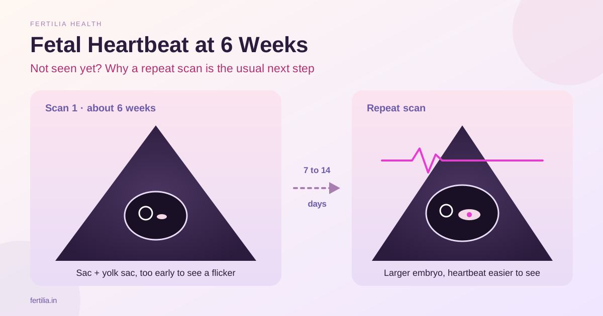
In early pregnancy, structures appear on scan in a set order. First the gestational sac, the small fluid-filled pocket in the uterus. Then the yolk sac, a tiny bright ring inside it. Then the fetal pole, the first visible form of the embryo, lying beside the yolk sac. The heartbeat shows as a flicker within the fetal pole once the embryo is large enough for the probe to pick it up.
On a transvaginal scan, that flicker typically becomes visible from around six to six and a half weeks, counted from the first day of your last period. At exactly six weeks, it is common to see a sac and a yolk sac, perhaps a very small fetal pole, and no flicker yet. Our radiologist’s guide to what an early scan shows from 6 to 8 weeks walks through that sequence week by week.
The type of scan matters too. A scan through the abdomen at six weeks sees much less than a transvaginal scan, because the probe sits several centimetres further from a uterus that is still small. If your scan was abdominal, a heartbeat that was not seen tells us even less. Our guide to why the transvaginal scan gives sharper early images explains the difference.
Why “6 Weeks” May Be 5 Weeks for Your Baby
Pregnancy weeks are counted from the first day of your last period, not from conception. That system assumes you ovulated on about day 14 of a 28-day cycle. Many women do not.
A prospective study that tracked ovulation in 696 cycles found that the fertile window fell entirely within days 10 to 17 of the cycle in only about 30% of women. Most reached it earlier, others much later, and the authors noted that timing can be highly unpredictable even when cycles are usually regular (Wilcox AJ et al., BMJ, 2000, PMID 11082086). If you ovulated on day 20 instead of day 14, a scan “at six weeks” is looking at a pregnancy that is effectively five weeks and a day or two old. At that stage, no heartbeat is the expected finding.
Implantation timing adds a little more variation. In a study of 221 women trying to conceive, implantation in pregnancies that continued happened between 6 and 12 days after ovulation, most often on day 8, 9 or 10 (Wilcox AJ et al., N Engl J Med, 1999, PMID 10362823). A few days here and a few days there add up quickly when the structures we are looking for appear within days of each other.
This is why the gap between your period date and your scan is so common if you:
- Have irregular or long cycles, including with PCOS
- Conceived soon after stopping the pill, or while breastfeeding
- Are not certain of your last period date
- Had bleeding in early pregnancy that you mistook for a period
If you conceived through IUI or IVF, your dates are far more precise, because they are counted from a known insemination or transfer date. Even then, your fertility team will usually repeat the scan before drawing any conclusion when the heartbeat is not seen at the first look.
What the Report Phrases Mean
Early scan reports use a compact language. These are the phrases I am most often asked about:
| What the report says | What it means |
|---|---|
| Single intrauterine gestational sac | The pregnancy sac is inside the uterus, in the right place |
| Yolk sac seen | A true pregnancy sac with early development under way |
| MSD (mean sac diameter) | The average size of the sac, in millimetres |
| Fetal pole seen / not seen | Whether the early embryo is visible yet |
| CRL (crown-rump length) | The length of the embryo, used for dating once it is visible |
| Cardiac activity not seen / FHR not seen | No flicker was seen on this scan |
| Pregnancy of uncertain viability | A sac or embryo is seen, but it is too early to say how things will progress |
| Suggest repeat scan after 7 to 14 days | The plan: wait, then look again |
“Pregnancy of uncertain viability” sounds frightening on paper. In practice, it is a description of timing: the pregnancy is in the uterus, and it is too early on the scan to know more. Many of these pregnancies go on to show a heartbeat at the next scan.
💜 Got a 6-week scan report with “cardiac activity not seen” and not sure what it means? Message Dr. Suganya on WhatsApp to book a ₹399 video consultation. Share your report and your period dates, and we can go through the numbers together and plan your next scan.
Why the Next Step Is a Repeat Scan
When a heartbeat is not seen and the embryo or sac is still small, the standard next step is a repeat transvaginal scan after a set interval, usually 7 to 14 days. That gap is chosen on purpose. It gives the pregnancy enough time to grow past the size at which each structure becomes visible, so the second scan can give a clear answer instead of another “too early to say”.
The caution is built on evidence. In 2011, a study of 1,060 women whose early scans were uncertain looked at the size cut-offs that some guidelines then used to diagnose miscarriage. With some of the smaller cut-offs, a proportion of pregnancies labelled as failing turned out to be healthy at the 11 to 14 week scan. The authors called some of those definitions potentially unsafe and suggested larger cut-offs of more than 25 mm for the sac and more than 7 mm for the embryo (Abdallah Y et al., Ultrasound Obstet Gynecol, 2011, PMID 21997898).
Guidance changed after that. The Society of Radiologists in Ultrasound published consensus criteria in 2013 that require either a clearly large sac or embryo, or a defined interval between two scans, before a pregnancy is called non-viable (Doubilet PM et al., N Engl J Med, 2013, PMID 24106937). A large UK study of 2,845 women later confirmed that those size cut-offs were appropriate and not too conservative (Preisler J et al., BMJ, 2015, PMID 26400869).
So when your doctor asks you to wait, they are following criteria written specifically to protect pregnancies that are healthy but early.
What Would Change the Plan
It helps to know what the repeat scan is looking for, so the second visit feels less like a test you might fail.
What most women hope to see, and often do: the embryo has grown, and a heartbeat is now visible. From here, your doctor will usually date the pregnancy by the embryo’s length (CRL) rather than your period date, and that may move your due date by a few days or a week. That is normal and does not mean anything is wrong.
When the picture needs a closer look: the 2015 UK study identified findings that reliably meant the pregnancy would not continue (Preisler J et al., BMJ, 2015, PMID 26400869). On a single scan, these were an empty sac with a mean diameter of 25 mm or more, or an embryo with a crown-rump length of 7 mm or more and no visible heart activity. Across two scans, they included an embryo without heart activity on both the first scan and a repeat scan seven or more days later, and a sac with no embryo whose size had not doubled after 14 days or more (when it started below 12 mm), or still showed no embryo heartbeat after seven days or more (when it started at 12 mm or above).
A small embryo with no heartbeat at six weeks does not meet any of these. That is why one early scan, on its own, is not used to decide anything.
If none of those findings is present but the pregnancy is still not clear, your doctor may suggest one more scan, or check a beta-hCG blood test alongside it. Our guide to beta-hCG levels and how they are read alongside the scan explains why a single number means less than the trend.
If no sac was seen in the uterus at all: this is a different situation from a sac without a heartbeat. It may simply be very early, but your doctor will want to follow your hCG levels closely until the pregnancy is located, to make sure it is not growing outside the uterus. Our post on ectopic pregnancy signs and what to expect explains why this follow-up matters.
When Not to Wait for the Repeat Scan
A heartbeat that is not yet seen is not an emergency in itself, and you do not need bed rest or to stop your normal day. Keep taking any folic acid or medicines your doctor has prescribed, and carry on with ordinary activity.
Please go to the nearest hospital straight away, without waiting for a call back or an appointment, if you have any of these. If you faint, collapse or feel too unwell to travel safely, call an ambulance:
- Heavy bright red bleeding, soaking a pad in an hour or less
- Severe pain in the lower tummy, especially on one side
- Pain in the tip of your shoulder
- Feeling faint, dizzy or very unwell
- Fever, or passing clots or tissue
Light spotting or brown discharge without pain is common in early pregnancy and does not usually need emergency attendance. Do let your doctor know the same day, so they can advise whether it can safely wait for your planned scan. This matters most if your scan has not yet clearly shown the pregnancy inside the uterus. Our guide to threatened miscarriage and what happens next explains what bleeding in the first weeks can and cannot tell you.
Getting Through the Wait
Seven to fourteen days can feel very long when you are waiting for a flicker on a screen. A few things help:
- Write down your questions now, while they are fresh, and take them to the next scan.
- Ask for a transvaginal scan at the repeat visit if your first one was abdominal, and keep the same centre if you can, so the measurements are easy to compare.
- Bring your first report. The repeat scan is read against it, and growth between the two is what tells the story.
- Go easy on searching. Most of what comes up is written for a different week, a different scan or a different situation.
- Notice your symptoms without testing them. Nausea and breast tenderness come and go in early pregnancy, and a quieter day does not tell you what the scan will show.
If this pregnancy follows a loss, the waiting can stir up much more. Our psychiatrist’s guide to pregnancy after miscarriage and the anxiety around scans has practical ways to manage those days.
If the repeat scan does bring difficult news, you will not be rushed into a decision. Our post on missed miscarriage and the options that follow explains each choice, and Fertilia’s free miscarriage support guide is there for the weeks after, for you and your partner.
What It Is Called in Hindi and Tamil
Many women search for this in their own language, or in a mix of English and Hindi or Tamil, such as “6 week me heartbeat nahi aayi”.
| Language | Term | Meaning |
|---|---|---|
| Hindi | बच्चे की धड़कन (bachche ki dhadkan) | The baby’s heartbeat |
| Tamil | குழந்தையின் இதயத் துடிப்பு (kuzhandhaiyin idhaya thudippu) | The baby’s heartbeat |
On scan reports in India, the English terms “cardiac activity”, “FHR” (fetal heart rate) and “fetal pole” are used across every language region.
Frequently Asked Questions
Is it normal not to see a heartbeat at 6 weeks?
Yes, it is common. On a transvaginal scan, the heartbeat usually becomes visible from around six to six and a half weeks, and if you ovulated later than day 14, your pregnancy may be closer to five weeks. A sac with a yolk sac, or a small embryo without a flicker yet, is a usual finding at this stage, and the standard step is a repeat scan in 7 to 14 days.
6 week me heartbeat nahi aayi, kya yeh normal hai?
Often, yes. At six weeks the heartbeat may simply be too small to see, especially if your period dates do not match when you ovulated. Doctors do not draw conclusions from one early scan when the embryo is small; they repeat the scan after 7 to 14 days to see how the pregnancy has grown.
When should the repeat scan be done?
Usually 7 to 14 days after the first scan, as your doctor advises. A shorter gap can give another unclear result, because the pregnancy has not had time to grow past the size where the heartbeat becomes visible. If you develop heavy bleeding or severe pain before then, do not wait for the planned date.
Can my dates be wrong if my cycles are regular?
Yes. Even women with regular cycles do not always ovulate on day 14. In one prospective study, only about 30% of women had their fertile window entirely within days 10 to 17 of the cycle (Wilcox AJ et al., BMJ, 2000, PMID 11082086). Once the embryo is visible, its length (CRL) gives a more accurate date than your period.
What size of embryo should show a heartbeat?
In the consensus criteria used by radiologists, an embryo of 7 mm or more in crown-rump length without a heartbeat is one of the findings that indicates the pregnancy will not continue (Doubilet PM et al., N Engl J Med, 2013, PMID 24106937). Below that size, the absence of a heartbeat on its own is not enough to decide, which is why a repeat scan is planned.
Should I rest in bed until the next scan?
No. Carrying on with your normal daily activity is fine, and there is no good evidence that bed rest changes what the next scan will show. Keep taking any prescribed folic acid or medicines, and go to the nearest hospital straight away if you have heavy bleeding, severe one-sided pain, shoulder-tip pain or faintness.
Does no heartbeat at 6 weeks mean I am going to miscarry?
Not on its own. A single early scan with no heartbeat, while the embryo or sac is still small, does not predict loss by itself. The criteria for diagnosing miscarriage were deliberately set to avoid that kind of early conclusion, and many of these pregnancies show a heartbeat at the next scan.
You Do Not Have to Read This Report Alone
💜 Waiting for your repeat scan and want someone to look at your report with you? Message Dr. Suganya on WhatsApp to book a ₹399 video consultation, available online across India. Bring your scan report and period dates, and we will go through what was seen, what the next scan is looking for and which questions to ask, alongside the care you are already receiving from your own doctor.