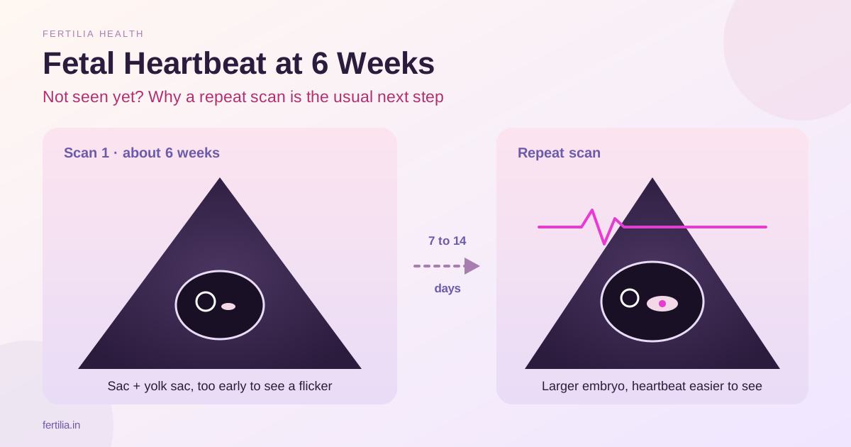
The scan room is quiet. The sonographer moves the probe and pauses. “I can see a gestational sac,” she says, “but I cannot see the heartbeat yet. Come back in ten days.”
You walk out with a printout showing two measurements, no image that looks like a baby, and a question looping in your head: does “no heartbeat” mean there is no heartbeat, or does it mean the scan was too early to find one?
It usually means the second. The timeline of early pregnancy development is narrow and predictable. What the scan finds at exactly six weeks, at seven weeks, and at eight weeks follows a precise sequence. Most of the time, when things look “sparse” on an early scan, the embryo is simply a week behind the point at which each structure becomes detectable.
I am a radiologist. I report these scans every working day. Here is the sequence, in plain language, so that the next scan report makes sense the first time you read it.
What this post covers:
- How early pregnancy develops week by week and when each structure appears on scan
- What a 6-week scan typically shows (and what it does not yet show)
- What changes at 7 and 8 weeks
- What the measurements on your report mean: MSD, CRL, FHR
- Why “too early to see” is not the same as “something is wrong”
- When a repeat scan is appropriate and why that is standard practice
- How the early scan connects to your beta-hCG level
The Sequence: What Develops and When
Early pregnancy has a precise developmental timetable. Ultrasound can only detect a structure once it has reached a size the probe can resolve. On a standard transvaginal scan, that threshold is roughly 2 to 3 millimetres.
| Week (from LMP) | What the embryo does | What TVS typically shows |
|---|---|---|
| 4 to 4.5 weeks | Implantation complete; gestational sac forms in uterine wall | Gestational sac (4–5 mm) in the uterine cavity |
| 5 to 5.5 weeks | Yolk sac forms inside the gestational sac | Yolk sac (3–5 mm); no fetal pole yet |
| 5.5 to 6 weeks | Fetal pole (embryo) emerges beside yolk sac | Fetal pole with possible early cardiac flicker |
| 6 to 7 weeks | Heart tube beats reliably; embryo doubles in size | Fetal pole with visible heartbeat; CRL 4–10 mm |
| 7 to 8 weeks | Limb buds forming; head becomes distinct | CRL 10–20 mm, clear cardiac activity, FHR 120–180 bpm |
| 8 weeks | Basic anatomy forming; amniotic cavity expanding | CRL around 20 mm, unmistakable heartbeat, yolk sac separating |
The numbers in this table are averages. Individual variation of five to seven days is normal, and gestational age calculated from the last menstrual period (LMP) can be out by that margin if cycles are irregular. A discrepancy of less than seven days between LMP-based dates and CRL-based dates is expected.
What a 6-Week Scan Shows
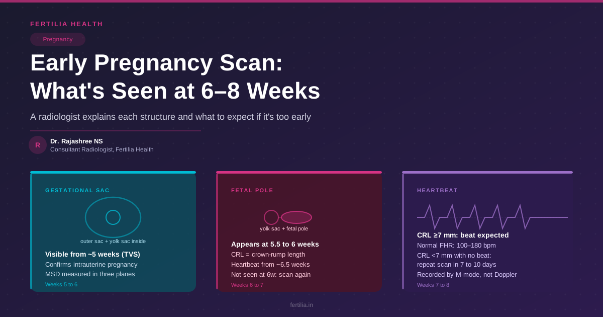
At exactly six weeks, a transvaginal scan should reliably show two things: a gestational sac in the uterine cavity and a yolk sac inside it.
The gestational sac is a small round or oval anechoic (fluid-filled, dark on screen) structure with a bright echogenic rim. The radiologist measures its mean sac diameter (MSD): the average of three measurements taken in different planes. At six weeks, this is typically between 16 and 24 mm.
The yolk sac appears as a small bright ring inside the gestational sac, usually between 3 and 6 mm in diameter. It is the embryo’s earliest nutrition source, before the placenta takes over. Seeing a yolk sac has two important meanings: it confirms the sac is a true gestational sac (not a pseudo-sac, which can occur in ectopic pregnancy), and it tells the radiologist that development is proceeding in the right direction.
What a 6-week scan does not reliably show is a fetal pole, and even less reliably, a heartbeat. The fetal pole becomes consistently visible from around 5.5 to 6 weeks by TVS. The heartbeat typically appears from 6 to 6.5 weeks. If your scan is done at exactly six weeks, or if your LMP-based date is slightly off and the embryo is effectively at 5.5 weeks, neither structure may yet be visible. That finding is not the same as an abnormal pregnancy.
At 7 Weeks: The Fetal Pole and Heartbeat
By seven weeks, the scan picture changes substantially. The fetal pole (the first distinct embryonic structure) is now consistently visible alongside the yolk sac. The radiologist measures the crown-rump length (CRL): the straight-line distance from the top of the embryo to its base. At seven weeks, CRL is typically between 8 and 12 mm.
The heartbeat should be visible at this stage. On ultrasound, early cardiac activity appears as a rapid flutter or flicker within the fetal pole. The radiologist records the fetal heart rate (FHR) in beats per minute using M-mode (a motion-sensitive line through the embryo). Between 6 and 7 weeks, a normal FHR is approximately 100 to 160 beats per minute, rising toward 150 to 180 bpm by eight weeks.
A heartbeat confirmed at 7 weeks on a good-quality TVS is reassuring. Published evidence shows that once cardiac activity is confirmed in the first trimester, the ongoing risk of loss is substantially lower than at earlier, undifferentiated stages (Doubilet et al. 2013, NEJM, PMID 24106937).
At 8 Weeks: A Clearer Picture
By eight weeks, the embryo has grown considerably. CRL is typically between 16 and 22 mm. The scan gives a much clearer image: the head is becoming distinct from the body, the amniotic cavity is expanding, the yolk sac may be separating away as this happens, and the heartbeat is brisk and unmistakable.
The 8-week scan is also when the radiologist can begin to assess the uterus and ovaries more thoroughly. The corpus luteum cyst on the ovary (which produces progesterone to support the pregnancy in early weeks) is often visible and is a normal finding. Fibroids, if present, are noted and measured. Any incidental findings on the ovaries are documented. These additional findings go into the report but do not usually require immediate action at this stage.
Understanding Your Report: Three Key Measurements
Most early pregnancy scan reports carry three core measurements. Here is what they mean:
Mean Sac Diameter (MSD): The gestational sac measured in three planes, with the results averaged. Used mainly when no fetal pole is yet visible. As a rough guide: an MSD of 25 mm or more without a visible embryo is one of the findings that prompts further investigation (Doubilet et al. 2013, PMID 24106937).
Crown-Rump Length (CRL): Measured once the fetal pole is visible. This is the most accurate dating tool in the first trimester, considerably more reliable than LMP-based dating when there is any doubt about cycle length. ISUOG practice guidelines recommend dating from CRL between 6 and 13 weeks 6 days whenever possible, with a margin of plus or minus 5 to 7 days (Salomon et al. 2019, Ultrasound Obstet Gynecol, PMID 31169958). A CRL of 7 mm or more without a heartbeat is the threshold at which the absence of cardiac activity becomes clinically significant; below 7 mm, the absence of a heartbeat alone is not sufficient grounds to draw conclusions.
Fetal Heart Rate (FHR): Recorded in beats per minute using M-mode. A rate between 100 and 180 bpm in the 6 to 8-week window is normal. Rates below 90 bpm at 7 weeks, or a heart rate that slows between two consecutive scans, are findings that a doctor will want to track closely. FHR cannot be measured accurately using colour Doppler at this stage; M-mode is the correct method.
Why “Too Early” Does Not Mean “Something Is Wrong”
This is the part of the conversation that matters most when a scan comes back without a heartbeat or without a fetal pole.
When the embryo is a genuine intrauterine pregnancy but the scan was done a few days before the structure reached detectable size, the scan looks “empty” or “sparse.” A gestational sac without a yolk sac at 5 weeks looks alarming; the same sac with a yolk sac one week later is completely normal. A fetal pole without cardiac activity at 6 weeks can be normal; the same embryo with a heartbeat at 6.5 weeks is reassuring.
The published criteria for diagnosing a non-viable pregnancy are deliberately conservative. They exist precisely to prevent incorrect diagnoses when the scan was simply early. The Society of Radiologists in Ultrasound consensus (Doubilet et al. 2013, PMID 24106937) specifies that no conclusion about viability should be reached on a single early scan unless the embryo is at or above the CRL threshold at which findings become definitive. Before any such conclusion is drawn, both the embryo size and the time elapsed since the first scan are considered.
If you are asked to return in 7 to 10 days, that is not a sign that something has gone wrong. It is standard practice. The repeat scan is a more informative data point than a single early scan, and the radiologist is waiting for the developmental timeline to catch up before drawing any conclusion.
If you have questions about an early pregnancy scan report or are unsure what your numbers mean, Dr. Suganya Venkat’s team offers an online consultation where you can go through the findings in detail.
Chat with Dr. Suganya’s team on WhatsApp
TVS Versus an Abdominal Scan at 6–8 Weeks
Almost all early first-trimester scans are done transvaginally (TVS). The transvaginal probe sits much closer to the uterus than an abdominal probe, which means it produces a sharper image at a smaller embryo size. A gestational sac that is 10 mm in diameter cannot be reliably imaged through an abdominal probe with the uterus sitting several centimetres away.
If you are given an abdominal scan at 6 weeks, the findings are more limited than a TVS. A gestational sac may be visible, but the yolk sac and fetal pole are frequently not seen by abdominal scan until 7 to 8 weeks. The absence of a fetal pole on an abdominal scan at 6 weeks is, therefore, not informative in the same way as the same finding on a TVS. For an explanation of how the transvaginal scan works and why it is preferred in early pregnancy, see Transvaginal Scan (TVS): What It Shows & Why It’s Done.
The Early Scan and Your Beta-hCG Level
Ultrasound findings in early pregnancy are closely linked to beta-hCG levels. Beta-hCG is the hormone produced by the developing pregnancy; it rises rapidly in the first weeks, typically doubling every 48 to 72 hours between 4 and 8 weeks.
A general principle (not a rigid threshold, as individual variation is wide): a gestational sac is typically visible on TVS when beta-hCG is above approximately 1,500 to 2,000 mIU/mL. A yolk sac is expected when levels are higher still. These are population-level guides, not individual cut-offs, and are never used in isolation.
If your beta-hCG levels are rising appropriately but your scan shows only a gestational sac, the most likely explanation is that the pregnancy is genuine but slightly earlier than the LMP-based date suggests. A repeat scan in one week, at which point beta-hCG will be higher and the embryo larger, usually resolves the question. For a detailed guide to what your beta-hCG numbers mean and how they are interpreted alongside scan findings, see Beta hCG Levels by Week: Normal Range & What It Means.
What to Ask at Your Early Scan
Bringing a few specific questions to the scan helps you understand the report while the findings are still being reviewed:
- What is the mean sac diameter, and is it consistent with my dates?
- Can you see a yolk sac inside the gestational sac?
- Can you see a fetal pole? If yes, what is the CRL?
- Is there cardiac activity?
- Is the gestational sac inside the uterine cavity?
- Is there anything to note on the ovaries?
These questions are entirely appropriate to ask. Most sonographers and radiologists will be happy to walk through them.
When You Are Referred for the NT Scan at 11–13 Weeks
If the 6 to 8-week scan goes well, the next milestone scan in India is the NT scan (nuchal translucency), done between 11 weeks and 13 weeks 6 days. The NT scan has a different focus: it is part of the combined first-trimester screening for chromosomal conditions. For a full explanation of what nuchal translucency measures and what the result means in practice, see NT Scan (Nuchal Translucency): What the Measurement Means.
The early 6 to 8-week scan and the NT scan serve different purposes. The early scan answers: is this a viable, intrauterine pregnancy? The NT scan answers: what is the risk of certain chromosomal conditions in this particular pregnancy?
A Note on First-Trimester Uncertainty
The weeks between a positive pregnancy test and the NT scan are, for many women, the most uncertain of the entire pregnancy. The embryo is small, the scan findings are limited, and every number on the report carries weight.
If you have had a previous pregnancy loss, that uncertainty is amplified further. It is entirely reasonable to want a scan at 6 weeks, another at 7 weeks, and another at 8 weeks. Most doctors will support this approach if the early scan is inconclusive or if there is a history of loss.
What I would ask you to hold on to is the developmental logic: each structure appears on its schedule. A scan done one week too early will look different from a scan done at the right moment, and that difference is almost always just the passage of seven days.
For a full overview of what happens week by week across the first trimester, including symptoms, tests, and what to watch for, see First Trimester: Symptoms, Tests & What to Expect.
FAQ
What should a 6-week scan show on a transvaginal scan?
A 6-week TVS should show a gestational sac in the uterine cavity and, in most cases, a yolk sac within it. A fetal pole may or may not be visible at exactly 6 weeks. A heartbeat is sometimes seen from 6 to 6.5 weeks, but its absence at this stage is not diagnostic of a problem. If only the sac and yolk sac are visible, a repeat scan in 7 to 10 days is the standard next step.
Why couldn’t the doctor see a heartbeat at my 6-week scan?
Cardiac activity typically becomes visible on TVS between 6 and 6.5 weeks of gestational age. If your scan was done right at 6 weeks, or if your cycle is slightly longer than average and the embryo is effectively at 5.5 weeks, the heartbeat may simply not yet be detectable. This is not a sign that the heart is not forming. A repeat scan at 7 weeks usually resolves this.
What is the yolk sac, and what does it do?
The yolk sac is a small bright ring visible inside the gestational sac from around 5 to 5.5 weeks. It provides nutrition and support to the early embryo before the placenta is fully formed. Seeing a yolk sac confirms the sac is a true gestational sac and that the right structures are developing. Its normal diameter is between 3 and 6 mm. An unusually large or irregular yolk sac may be noted in the report for monitoring.
What is a fetal pole?
The fetal pole is the first visible form of the embryo, appearing at around 5.5 to 6 weeks as a small thickening next to the yolk sac. The radiologist measures it as the crown-rump length (CRL). Once visible, the fetal pole is the structure monitored for cardiac activity and for size, which determines gestational age more accurately than the LMP date.
My scan showed an empty gestational sac with no yolk sac. What does that mean?
An empty gestational sac with no yolk sac may simply mean the scan was done before 5 weeks, when the yolk sac has not yet become visible. If the mean sac diameter is 25 mm or more without a visible embryo or yolk sac, that combination meets one of the criteria that warrants further assessment. The radiologist will note this in the report and recommend either a follow-up scan or a repeat beta-hCG level.
How accurate is the 6 to 8-week scan for dating my pregnancy?
Crown-rump length measured between 6 and 14 weeks is the most accurate method of pregnancy dating currently available. The margin of error is plus or minus 5 to 7 days. If the difference between your LMP-based date and the scan-based date is less than 7 days, the LMP date is usually kept. A discrepancy of 7 days or more means the CRL-based date takes precedence. Per ISUOG practice guidelines (Salomon et al. 2019, PMID 31169958), CRL-based dating is the preferred method when it is available within this window.
What measurements does the radiologist record on an early pregnancy scan?
The three core measurements are: mean sac diameter (MSD, the average of three sac dimensions, used before the fetal pole is visible), crown-rump length (CRL, the embryo’s length once visible), and fetal heart rate (FHR, in beats per minute, recorded by M-mode once cardiac activity is confirmed). The report also notes the location of the sac (intrauterine or otherwise), any findings in the myometrium, and the appearance of both ovaries.
If your early scan report has raised questions, or if you have had more than one inconclusive scan and are not sure what it means for your pregnancy, a detailed online consultation with Dr. Suganya Venkat’s team can help you understand the findings clearly.