A woman comes in for the preconception health check recommended before a fertility program starts. Her gynaecologist adds a thyroid ultrasound to the panel. The report arrives the next morning: “right thyroid lobe nodule, 1.2 x 0.9 cm, solid, hypoechoic, TIRADS 3, no suspicious margins or calcification noted.” She searches TIRADS 3 on her phone. Somewhere in the second page of results she reads the word “malignancy.” She calls the clinic asking whether she has thyroid cancer.
She does not. TIRADS 3 represents a mildly suspicious nodule with a malignancy risk of roughly 2 percent, and at 0.9 cm it does not yet meet the size threshold where a tissue sample is recommended. But the report she is holding does not say any of that. It was written for the clinician who ordered the scan, using shorthand that clinicians read fluently and most patients find frightening without context.
I am a radiologist, and thyroid nodules are one of the most common incidental findings I document in reports. They are also, in my experience, among the most consistently misread by patients who try to interpret the printout themselves. This guide translates the standard thyroid nodule ultrasound report into plain language, explains the TIRADS scoring system category by category, and describes when a follow-up or procedure is genuinely needed.
You may have come across my earlier guides to reading a follicular study report or an ovarian cyst report. The approach here is the same: not a general article about thyroid health, but a working guide to the specific language of the scan report you are holding.
What this post covers:
- How common thyroid nodules are, and why most are found by accident
- What the ultrasound is measuring, and what a nodule actually is
- The TIRADS classification: every category, what it means, what it does not mean
- How to read the other terms in your report
- When FNAC is recommended, and the Bethesda system for cytology results
- What a thyroid nodule means for fertility and pregnancy planning
How common are thyroid nodules?
The first thing to know before reading the rest of your report is a number: more than half of adults who undergo a high-resolution thyroid ultrasound will have at least one nodule detected (Haugen et al., 2016, ATA Management Guidelines, PMID 26462967). The majority of these are found incidentally during a neck scan ordered for something unrelated, or as part of a general preconception or health checkup.
Most thyroid nodules are benign. Across populations of nodules that are biopsied (a sample already selected toward higher suspicion), malignancy rates range from 7 to 15 percent. Among all detected nodules, including the many that are simply watched and never sampled, the proportion with any clinical significance is considerably smaller.
India has had regions of historical iodine deficiency, particularly in inland areas where coastal fish consumption is low. Iodine deficiency promotes thyroid nodule formation and goitre. The national iodisation programme has improved coverage significantly in recent decades, but this background is worth knowing when your scan was done in a state with historically lower iodine intake.
What the thyroid ultrasound is measuring
The thyroid is a butterfly-shaped gland at the front of the neck, below the voice box. It has two lobes, one on each side, joined by a narrow bridge called the isthmus. A thyroid ultrasound uses high-frequency sound waves to image the gland. There is no radiation and no injection of contrast.
A nodule is a discrete region within the thyroid that reflects sound waves differently from the surrounding tissue. It is not inherently dangerous, and it is not a tumour until a tissue sample proves otherwise. What the radiologist is doing, in practical terms, is characterising that difference: is the nodule solid or fluid-filled? Is it darker or brighter than surrounding tissue? Does it have an unusual shape, irregular edges, or certain types of calcification? These features, taken together, generate the TIRADS score.
The TIRADS classification: what each category means
TIRADS stands for Thyroid Imaging Reporting and Data System. The version most radiologists in India now use is the ACR TI-RADS, published by the American College of Radiology (Tessler et al., J Am Coll Radiol, 2017;14(5):587-595, PMID 28372962). It assigns points based on five features of the nodule, then places it in a category.
The five scored features are:
- Composition: what the nodule is made of (pure cyst, spongiform, mixed, or solid)
- Echogenicity: how bright or dark the nodule appears relative to thyroid tissue
- Shape: wider-than-tall (lower risk) versus taller-than-wide (higher risk)
- Margin: smooth, ill-defined, lobulated or irregular, or extending beyond the thyroid capsule
- Echogenic foci: types of bright spots or calcification within the nodule
The categories:
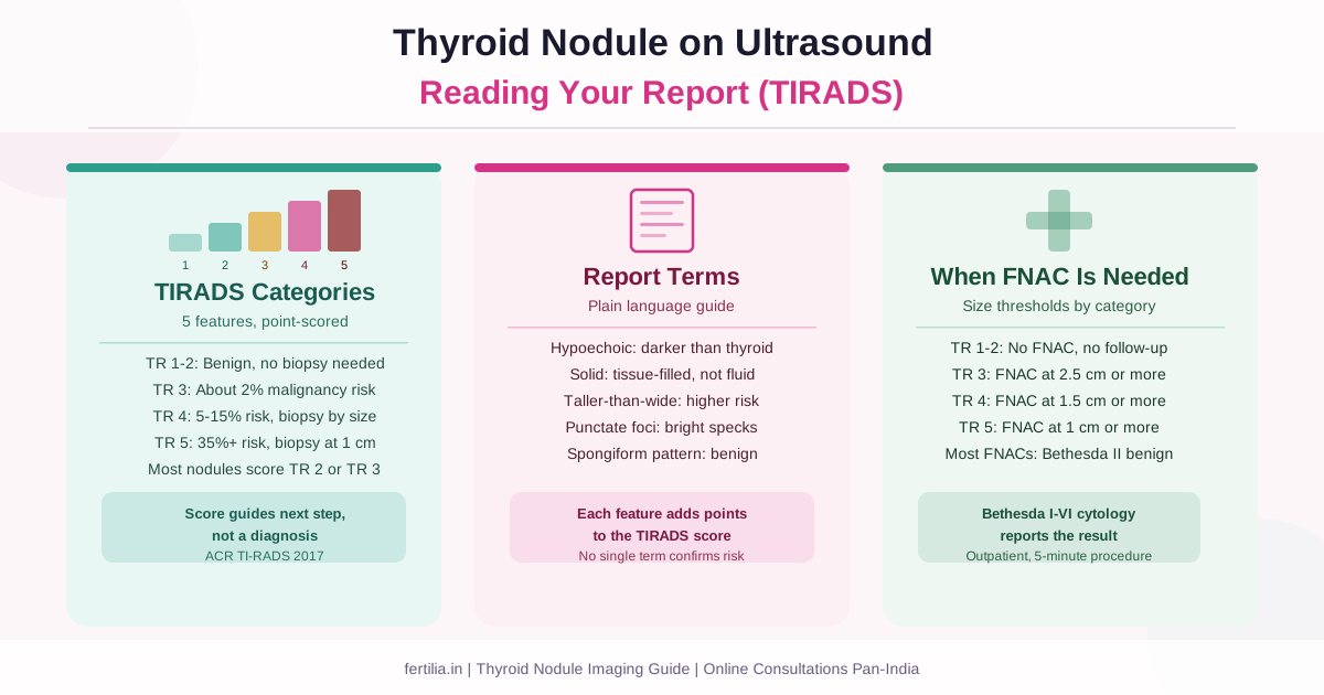
TR 1 (TIRADS 1): Normal thyroid. No nodule is present, or the finding is entirely normal tissue. No action needed.
TR 2 (TIRADS 2): Benign. Pure cysts with no solid component score here. The malignancy risk is effectively zero. No follow-up scan or biopsy is needed.
TR 3 (TIRADS 3): Mildly suspicious. Malignancy risk approximately 2 percent. The majority of incidentally found nodules sit in this category. Follow-up scanning is recommended when the nodule reaches 1.5 cm; biopsy when it reaches 2.5 cm or larger.
TR 4 (TIRADS 4): Moderately suspicious. Malignancy risk approximately 5 to 15 percent. Biopsy is recommended at 1.5 cm or larger, and follow-up scanning from 1 cm onward.
TR 5 (TIRADS 5): Highly suspicious. Malignancy risk approximately 35 percent or higher. Biopsy is recommended at 1 cm, with follow-up from 0.5 cm if the nodule does not yet meet the biopsy threshold. This is the category where the radiologist’s report typically includes a specific recommendation for clinical review.
Two things to hold on to. First, TIRADS 5 is not a diagnosis of cancer. It is a scoring system that guides the decision about whether to biopsy. The biopsy is what tells you whether cancer is present, and even among nodules that are biopsied, most results are benign. Second, most nodules in practice score TIRADS 2 or 3. TIRADS 4 and 5 nodules are less common in a general population, more common in a clinic that sees women sent specifically because of a finding.
Reading the other terms on your report
Hypoechoic: The nodule is darker than the surrounding thyroid tissue. This contributes to the TIRADS score because hypoechoic nodules carry a slightly higher association with malignancy than those that match the surrounding tissue or appear brighter. It does not mean cancer on its own.
Isoechoic: The nodule matches the brightness of surrounding tissue. Lower risk category on the echogenicity scale.
Hyperechoic: Brighter than surrounding tissue. One of the lower-risk echogenicity patterns.
Solid vs spongiform vs cystic: A solid nodule is filled entirely with tissue and carries the highest echogenicity score. A spongiform nodule has a sponge-like appearance, with tiny fluid pockets making up more than half its volume; this pattern is benign and scores zero on composition. A cystic nodule is fluid-filled; pure cysts are TIRADS 2.
Macrocalcification: A large, coarse calcification that casts an acoustic shadow behind it on the scan. Commonly seen in old benign nodules with degenerative change. Not a sign of cancer by itself.
Peripheral calcification: Calcification along the outer rim of the nodule, sometimes described as eggshell calcification. Associated with benign findings in most cases.
Punctate echogenic foci: Small, bright specks within a solid nodule. There are two types. Comet-tail artefact foci (with a reverberating bright tail behind the speck) are benign, caused by colloid. Non-comet-tail punctate echogenic foci are the highest-scoring finding in the ACR TIRADS system, and are associated in some cases with papillary thyroid cancer. Many nodules with these foci are still benign when biopsied. The finding raises the TIRADS score and may bring the nodule into a follow-up or biopsy recommendation, but it is not a diagnosis.
Vascularity (colour Doppler): Some reports add colour Doppler imaging to assess blood flow within the nodule. Blood flow patterns are not part of the ACR 2017 TIRADS score (they were in older classification systems), so this line on your report is additional clinical information rather than a score-determining feature.
If your report was written using an older TIRADS classification, the category numbers may differ slightly from the ACR 2017 system described here. The system used should be specified; if not, ask the radiologist who signed your report or the clinician who ordered it.
When FNAC is recommended
FNAC (fine needle aspiration cytology, or fine needle aspiration biopsy) is the procedure used to sample a thyroid nodule. A radiologist guides a thin needle into the nodule under real-time ultrasound, draws out a small number of cells, and sends these to a pathologist who examines them under a microscope. The procedure takes about five minutes, uses a very fine needle, and causes discomfort similar to a blood draw for most people. No preparation or fasting is needed. It is done on an outpatient basis.
Whether FNAC is recommended depends on the TIRADS category and the nodule’s size. The ACR 2017 thresholds:
- TR 1 or TR 2: No FNAC, no follow-up imaging
- TR 3: Follow-up scan if 1.5 cm or larger; FNAC if 2.5 cm or larger
- TR 4: Follow-up scan if 1 cm or larger; FNAC if 1.5 cm or larger
- TR 5: Follow-up scan if 0.5 cm or larger; FNAC if 1 cm or larger
A TR 4 nodule smaller than 1.5 cm does not need FNAC; it is placed on a follow-up schedule. This is intentional. Very small nodules, even if they score as moderately suspicious on imaging, are slow-growing in most cases and rarely change management in a meaningful way at a small size.
When FNAC is done, the result is reported using the Bethesda system (Cibas and Ali, Thyroid, 2017;27(11):1341-1346, PMID 29091573), which has six categories:
- Bethesda I: Non-diagnostic. The sample did not yield enough cells for interpretation. Repeat FNAC is usually recommended.
- Bethesda II: Benign. The most common result. Includes colloid nodules, thyroiditis, simple cysts.
- Bethesda III: Atypia of undetermined significance. A grey zone; often leads to a repeat FNAC or molecular testing.
- Bethesda IV: Follicular neoplasm. Imaging and cytology cannot distinguish a follicular adenoma from follicular carcinoma, so surgical removal is usually recommended for a definitive diagnosis.
- Bethesda V: Suspicious for malignancy.
- Bethesda VI: Malignant.
Among all FNACs performed, Bethesda II (benign) remains the most common outcome, including among nodules sampled because they scored higher on TIRADS.
In India, FNAC of the thyroid is available at most large diagnostic centres and at government teaching hospitals. The procedure is the same whether done at a private lab or a government hospital; the waiting times and costs differ.
Have you received a thyroid scan report with a TIRADS score and want to understand what it means alongside your fertility or preconception plan? Message us on WhatsApp and Dr. Suganya’s team can help you connect the imaging finding with the clinical picture.
Thyroid nodules and fertility: what the finding means for your plan
If you are reading this because you have a thyroid nodule and are planning a pregnancy, here is the practical answer: in most cases, the nodule itself is not the issue. What matters for fertility and pregnancy is thyroid function, which is measured by TSH, free T4, and where clinically indicated, anti-TPO antibodies. These are separate questions from the nodule.
A thyroid nodule found incidentally in a woman whose TSH is normal does not usually require treatment before trying to conceive. The nodule follow-up schedule (if one is recommended based on TIRADS and size) continues alongside the fertility workup, not instead of it.
If the nodule is a functioning nodule, meaning it is producing thyroid hormone autonomously, it can cause hyperthyroidism. This is considerably less common than the typical incidentally found non-functioning nodule, and a nuclear medicine thyroid scan (scintigraphy) can distinguish the two. Hyperthyroidism affects fertility and requires treatment, but this is a separate conversation from the standard nodule finding.
Pregnancy itself can cause mild growth of pre-existing nodules in some women, because high hCG levels in the first trimester stimulate TSH receptors. FNAC is safe in pregnancy if it becomes indicated. The assessment does not usually change because a woman is pregnant.
If you also have Hashimoto’s thyroiditis (autoimmune thyroid disease), nodules and Hashimoto’s frequently coexist. Our guide to Hashimoto’s and conception explains how the autoimmune component separately affects miscarriage risk and TSH targets, which is a distinct question from any structural nodule. For the broader picture of thyroid function and fertility, our thyroid and fertility guide covers TSH targets, T3/T4 ranges for conception, and the tests that are directly relevant.
If you have subclinical hypothyroidism alongside a nodule, the TSH management question is usually the one that directly affects the fertility timeline, not the nodule itself. The preconception checkup we recommend typically covers both the function tests and the imaging, so the clinical picture is assessed together. You can find an overview of the standard preconception panel at our preconception checkup resource.
Practical notes for the follow-up scan
Follow-up scan timing for TR 3 nodules: if the nodule is between 1.5 cm and 2.5 cm, the ACR recommends a repeat ultrasound at 1, 2, and 5 years. For TR 3 nodules smaller than 1.5 cm, no follow-up imaging is needed. For TR 4 nodules between 1 cm and 1.5 cm that do not yet meet the FNAC threshold, a repeat scan at 1, 2, 3, and 5 years is the recommended schedule.
If the nodule grows by 20 percent or more in two dimensions, or by 2 mm in solid components, at any follow-up scan, that is considered significant growth and typically prompts upgrading to FNAC regardless of whether it has reached the size threshold.
If you have a TIRADS 4 or 5 finding and the report does not include a clear recommendation, ask the ordering clinician or request a direct telephone review with the radiology department that issued the report. Most centres offer this.
Frequently asked questions
What does TIRADS mean on a thyroid ultrasound report?
TIRADS stands for Thyroid Imaging Reporting and Data System. It is a point-based scoring method used to classify how suspicious a thyroid nodule appears on ultrasound. The ACR TI-RADS 2017 version, most widely used in India now, scores five features of the nodule (composition, echogenicity, shape, margin, echogenic foci) and places it in a category from TR 1 (normal) to TR 5 (highly suspicious). Higher categories indicate that closer monitoring or a tissue sample is needed, not that cancer is confirmed.
My report says TIRADS 4. Does that mean I have cancer?
No. TIRADS 4 means the nodule scored as moderately suspicious on imaging features. Approximately 5 to 15 percent of TIRADS 4 nodules, when biopsied, turn out to be malignant. That means 85 to 95 percent are not. Whether FNAC is recommended also depends on the nodule’s size: a TIRADS 4 nodule smaller than 1.5 cm would typically go on a follow-up scan schedule rather than be biopsied immediately. Your treating doctor will advise on the next step.
Does a thyroid nodule affect my chances of getting pregnant?
In most cases, no. A nodule by itself does not affect fertility. What matters for conception is whether thyroid function is normal, measured by TSH and free T4. If your function tests are normal, the incidental nodule finding does not usually change your fertility management. Read more in our thyroid and fertility guide and the guide to conceiving with hypothyroidism.
What is FNAC for a thyroid nodule, and does it hurt?
FNAC (fine needle aspiration cytology) uses a thin needle, guided by real-time ultrasound, to draw out cells from the nodule for microscopic examination. The result is reported using the six-category Bethesda system. The most common outcome is Bethesda II (benign). The procedure takes about five minutes; most people describe the discomfort as similar to a routine blood draw. No fasting or preparation is needed, and it is done as an outpatient.
My report mentions “punctate echogenic foci” in the nodule. What does that mean?
Punctate echogenic foci are small, bright specks within a solid nodule. Comet-tail artefact foci (with a bright reverberating tail) are benign. Non-comet-tail punctate echogenic foci score the highest number of points in the TIRADS system because they are associated, in some cases, with papillary thyroid cancer. However, many nodules with these foci are benign on biopsy. The finding raises the TIRADS score and may move the nodule into a follow-up or FNAC recommendation, but it is not a diagnosis.
How often do I need a follow-up scan for a thyroid nodule?
This depends on the TIRADS category and size. TR 3 nodules between 1.5 cm and 2.5 cm: follow-up scans at 1, 2, and 5 years. TR 3 nodules under 1.5 cm: no follow-up needed. TR 4 nodules between 1 cm and 1.5 cm: follow-up at 1, 2, 3, and 5 years. If a nodule shows significant growth at any scheduled scan (20 percent or more in two dimensions, or 2 mm increase in solid component), that usually prompts biopsy regardless of size.
Thyroid scan report mein TIRADS kya hota hai, aur thyroid ki gaanth ka kya matlab hai?
TIRADS ek scoring system hai jisme radiologist thyroid ki gaanth (nodule) ko classify karta hai ultrasound par. Thyroid ki gaanth matlab hai gland ke andar ek alag tissue region, jo bahut aam hai. 50 percent se zyada logo mein high-resolution scan par koi na koi nodule milti hai. TIRADS 1 se 5 tak categories hain: TIRADS 2 benign hoti hai, TIRADS 3 thodi suspect (lagbhag 2% malignancy risk), TIRADS 4 moderate suspect (5-15%), TIRADS 5 zyada suspect (35% ya usse zyada). Iska matlab cancer nahi hota. FNAC (biopsy) tab ki jaati hai jab nodule ek certain size se bari ho, category ke hisaab se.
If you have a thyroid nodule on your report and want guidance on how it connects to your fertility workup or pregnancy plans, Dr. Suganya’s team is here to help. We work online, pan-India, via video call. Message us on WhatsApp to start a conversation.