A woman receives her pelvic ultrasound report and the first line reads “right adnexal cyst, 3.8 x 3.2 cm, anechoic, thin-walled, posterior acoustic enhancement noted.” Her scan was ordered for mild lower abdominal discomfort. She has no other symptoms. The radiologist has written a precise, technically accurate report. But to the woman holding the printout, those three lines mean nothing, and by the time she reaches her doctor the next morning, she has spent two hours reading worst-case scenarios online.
For more on this, read our guide on Thyroid Nodule on Ultrasound.
For more on this, read our guide on Transvaginal Scan (TVS). This sequence is very common. The language of an ultrasound report is designed for clinician-to-clinician communication. It is precise and useful in that context, but it was not written for the woman who carries it home.
I am a radiologist, and I write reports like this daily. This guide translates a standard ovarian cyst ultrasound report into plain language so that you can read your own printout, understand what each term is telling you, and arrive at your doctor’s appointment knowing what questions to ask.
What this post covers:
- What “adnexal cyst” means and why the report is written that way
- The single most important distinction: simple vs complex
- A plain-language glossary of the terms found in pelvic ultrasound reports
- How the most common cyst types appear on scan
- What cyst size means for management
- The O-RADS scoring system you may see on your report
- When a follow-up scan is appropriate, and what it is checking for
What does “adnexal” mean?
The word “adnexal” refers to the structures that sit alongside the uterus on each side: the ovaries, the fallopian tubes, and the surrounding ligaments and tissue. When a radiologist writes “right adnexal cyst,” it means a cyst was found on the right side, in the region of the right ovary or tube.
In most cases, an adnexal cyst on a pelvic ultrasound is an ovarian cyst. The ovary is the most common source. Occasionally, a cyst arises from the fallopian tube (a paraovarian or paratubal cyst) and sits close enough to the ovary that the two cannot be easily separated on the scan. In those cases, “adnexal” is the accurate term because the precise origin is unclear from imaging alone.
This is not a cause for concern. It simply describes anatomy.
The most important line in your report: simple or complex?
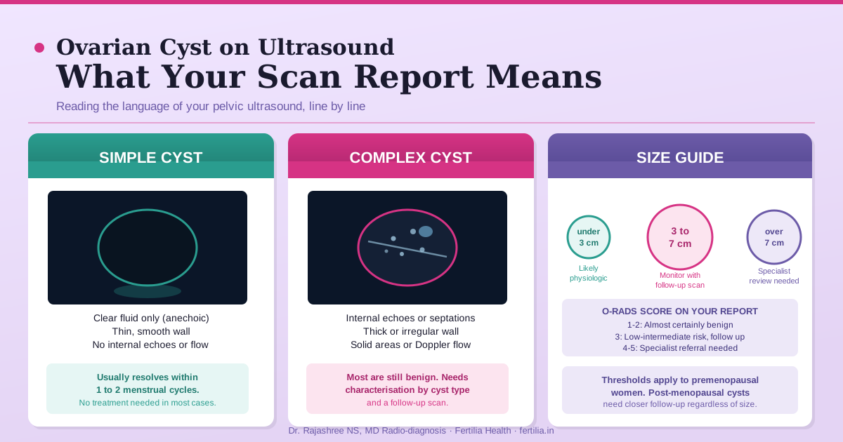
Before reading anything else, find this distinction in your report. A pelvic ultrasound report for an ovarian cyst will describe it as either a simple cyst or a complex cyst. This one word carries more clinical weight than the size measurement.
A simple cyst is:
- Anechoic: completely dark on scan, meaning it contains only clear fluid with no solid material
- Thin-walled: the cyst wall is smooth and fine, without thickening or irregularity
- Unilocular: one single chamber, no internal dividers
- Without internal echoes, solid components, or detectable blood flow within the cyst
A complex cyst is any cyst that deviates from those features. That might mean:
- Internal echoes (material suspended within the fluid, such as old blood or cellular debris)
- A thick or irregular wall
- Septations (internal dividing walls that create more than one compartment)
- Solid areas within or attached to the cyst
- Papillary projections (small finger-like growths on the inner wall)
- Detectable blood flow within the cyst on colour Doppler
A simple cyst is very rarely concerning in a woman of reproductive age. The vast majority are benign and functional: they form as a normal part of the ovarian cycle and resolve within one to three menstrual cycles. ACOG Practice Bulletin #174 (2016) on the evaluation and management of adnexal masses recommends expectant management for simple unilocular cysts under 5 cm in premenopausal women, meaning observation and a follow-up scan is usually all that is needed.
A complex cyst requires more attention, not because it is necessarily serious, but because the appearance needs context. The majority of complex cysts in reproductive-age women are still benign. A hemorrhagic cyst (where the follicle has bled) and an endometrioma (a cyst arising from endometriosis) are both “complex” on ultrasound and both almost always benign. The complexity tells the radiologist that the cyst contains more than clear fluid, and that further characterisation may be warranted.
Glossary: what each term in your report means
Here are the words most commonly found in a pelvic ultrasound report for an ovarian cyst. Read this section with your printout beside you.
Anechoic. No internal echoes. The cyst appears uniformly dark on the scan, meaning it contains only clear fluid. An anechoic cyst with a thin wall is the definition of a simple cyst.
Posterior acoustic enhancement. The area behind the cyst appears brighter than surrounding tissue. This happens because sound waves pass easily through fluid and bounce back more strongly beyond it. Posterior acoustic enhancement is a reassuring sign that a structure contains fluid, not solid material.
Internal echoes. The cyst contains material that reflects sound waves. This might be old blood, mucus, fat, or other tissue. It does not mean the cyst is malignant. It means the cyst is not a straightforward fluid-only structure and needs to be characterised more fully.
Septations. Thin internal walls that divide the cyst into two or more compartments. Fine, thin septations in an otherwise simple-looking cyst carry very low risk. Thick or irregular septations with blood flow prompt closer evaluation.
Papillary projections. Small, irregularly shaped solid components projecting inward from the cyst wall. In the context of the right patient age and hormonal history, these may be completely benign (as in an endometrioma with an adherent clot) or may require further imaging.
Vascularity / colour Doppler positive. Blood flow detected within the cyst wall or septations. Solid components with internal blood flow need specialist review. Detectable flow within a papillary projection is a finding that typically prompts referral. No internal flow within a cyst is reassuring.
Unilocular. One compartment only, no internal walls. A unilocular anechoic cyst is the simplest and least-concerning type of ovarian cyst.
Multilocular. More than one compartment. A multilocular cyst that is otherwise thin-walled and free of solid components can still be benign (a paraovarian cyst or a serous cystadenoma, for instance), but it requires follow-up.
Ground-glass echogenicity. Uniform, fine, low-level internal echoes that give the cyst a homogeneous, hazy appearance. This is the classic description of an endometrioma on ultrasound.
Echogenic focus with shadowing. A bright spot within the cyst that blocks sound waves behind it. This is a common finding in dermoid cysts (ovarian teratomas), where fat, hair, or calcification produces this appearance.
If you are looking at your scan report and are unsure what to do next, speaking with an OB-GYN who can review the full clinical picture is the most direct route to a clear answer.
Dr. Suganya Venkat is available online for a Rs. 399 video consultation, pan-India. You can share your scan report directly in the conversation and get a specific, evidence-based answer rather than a general one.
Common cyst types and how they appear on scan
Knowing the cyst type your report is describing is often the most useful piece of information. Here are the types a radiologist distinguishes on pelvic ultrasound:
Follicular cyst. The most common ovarian cyst in reproductive-age women. A follicle grows each month in preparation for ovulation. Occasionally it does not release the egg and continues to enlarge, forming a thin-walled, anechoic cyst. Almost always resolves within one to two menstrual cycles without treatment.
Corpus luteum cyst. After ovulation, the follicle collapses and forms the corpus luteum, a temporary hormone-producing structure. Sometimes it fills with fluid or blood, creating a cyst with an irregular, lacy inner wall and low-level internal echoes. These can be tender (particularly in the second half of the cycle), look complex on ultrasound, and yet resolve completely within six to eight weeks.
Hemorrhagic cyst. A functional cyst in which bleeding has occurred within the fluid. The internal echoes have a characteristic reticular (cobweb-like) or fishnet pattern. There may be no internal Doppler flow because the internal material is clot, not solid tissue. Hemorrhagic cysts resolve on their own in the large majority of cases. A follow-up scan at six to eight weeks confirms resolution.
Endometrioma. A cyst formed when endometrial tissue grows on the ovary. The fluid inside is old blood, giving the cyst a distinctive homogeneous ground-glass echogenicity: uniformly fine, low-level internal echoes throughout. The wall may be slightly irregular. An endometrioma does not resolve without treatment and is associated with endometriosis. If your report describes ground-glass echoes in a cyst in a woman with pelvic pain or painful periods, this is the cyst type your doctor will consider. Women with PCOS and endometriomas often have both conditions contributing to irregular cycles: the PCOS Symptom Reversal Program addresses the metabolic and hormonal layer that runs alongside endometriosis-related findings.
Dermoid cyst (mature cystic teratoma). A benign cyst containing hair, fat, and sometimes calcification, arising from germ cells in the ovary. On ultrasound it appears as a hyperechoic mass with acoustic shadowing, often with a Rokitansky nodule (a solid, echogenic nodule projecting into the cyst). Dermoids are almost always benign. They do not resolve on their own and may need surgical removal if they grow large or cause symptoms.
Paraovarian cyst. Arises from remnant tissue near the fallopian tube rather than from the ovary itself. Typically thin-walled, anechoic, and clearly separate from the ovary on careful scanning. Benign. Usually requires observation only.
What cyst size means for management
Size alone does not determine how a cyst is managed. The combination of size and appearance guides the decision. As a general framework, based on guidance from the American College of Obstetricians and Gynecologists (ACOG Practice Bulletin #174, 2016) and the American College of Radiology:
Under 3 cm. In a premenopausal woman, a simple anechoic cyst under 3 cm is most likely a normal follicle or a physiologic cyst. No follow-up imaging is typically needed.
3 to 5 cm. A simple cyst in this range is very likely benign. Your doctor may recommend a follow-up scan in one to two menstrual cycles to confirm resolution. A complex cyst in this size range should be characterised in more detail on the report, and a follow-up scan at six to eight weeks is standard.
5 to 7 cm. A simple cyst of 5 to 7 cm in a premenopausal woman is still very likely benign, but a follow-up ultrasound at 6 to 12 weeks is recommended to confirm stability or resolution. Complex cysts in this range need follow-up and possibly further characterisation with MRI, depending on their features.
Over 7 cm. Simple cysts over 7 cm are usually reviewed with MRI or specialist referral to characterise them fully. Surgical consultation may be appropriate depending on the appearance and the presence of symptoms. This does not mean surgery is necessary, only that the evaluation requires more information than ultrasound alone can provide.
These thresholds apply specifically to premenopausal women. In postmenopausal women, any cyst, regardless of size or simplicity, warrants more careful attention and a shorter follow-up interval, because the ovaries do not form functional cysts after menopause.
The O-RADS score: if it appears on your report
O-RADS stands for Ovarian-Adnexal Reporting and Data System. It is a structured scoring system developed by the American College of Radiology to standardise how radiologists describe and categorise adnexal findings, in the same way that BI-RADS is used for breast imaging. The system was published in 2020 (Andreotti RF et al., Radiology 2020; 294(1):168-185; PMID 31687921) and is now in use at many imaging centres in India.
If your report includes an O-RADS score, this is what each category means:
O-RADS 0. The scan is technically inadequate or incomplete. A repeat scan is needed.
O-RADS 1. Normal ovary. No cyst or mass. No follow-up needed.
O-RADS 2. Almost certainly benign. This category includes simple cysts under 10 cm, typical hemorrhagic cysts, dermoids with classic features, and endometriomas with classic features. Malignancy risk is less than 1%. Follow-up, if any, is based on size rather than concern about malignancy.
O-RADS 3. Low risk. Features that are not purely benign but still carry a malignancy risk under 10%. This includes smooth-walled multilocular cysts under 10 cm and cysts with mildly irregular features without solid components or Doppler flow. Usually followed up with repeat ultrasound or MRI in 8 to 12 weeks.
O-RADS 4. Intermediate risk. Malignancy risk between 10 and 50%. These findings warrant prompt referral for specialist evaluation.
O-RADS 5. High risk. Malignancy risk over 50%. Specialist referral is recommended urgently.
The majority of ovarian cysts found incidentally in reproductive-age women fall into the O-RADS 1 or O-RADS 2 category.
The follow-up scan: what it is checking
When a radiologist recommends a follow-up scan, the goal is usually one of three things: to confirm that a functional cyst (follicular or corpus luteum) has resolved, to check that a borderline cyst has not grown or changed in character, or to re-examine a cyst at a different point in the menstrual cycle when the appearance may be clearer.
A “follow-up scan in 6 to 8 weeks” is not a warning. It is the radiologist’s way of saying the cyst is not urgent but warrants one more look.
It is helpful to schedule the follow-up scan at a specific point in your cycle. A scan on Day 5 to 8 (counting Day 1 as the first day of bleeding) is often most informative: any functional cyst from the previous cycle has had time to resolve, and a new follicle has not yet grown large enough to be confusing. If your doctor has not specified a cycle-day, mention this when booking.
For women who are undergoing fertility investigations, an ovarian cyst finding on a pelvic scan is often interpreted alongside the antral follicle count, which counts small resting follicles as a measure of ovarian reserve. A large cyst in one cycle can obscure that count and make the AFC less accurate, which is another reason the follow-up scan timing matters.
What to tell your doctor after receiving this report
If your report describes a simple anechoic cyst under 5 cm, the conversation is usually brief: both the radiologist and your doctor expect it to resolve. The useful questions at that appointment are: when should I repeat the scan, should it be at a specific point in my cycle, and are there symptoms I should watch for in the meantime?
If your report describes a complex cyst, an endometrioma, or a dermoid, or if the O-RADS score is 3 or above, the conversation will be more specific to your situation: your symptoms, your hormonal history, whether you are trying to conceive, and what further imaging or referral is appropriate.
Seek assessment before the scheduled follow-up if you develop any of the following: sudden severe lower abdominal pain, fever, nausea with rapid-onset one-sided pain, or pain severe enough to interrupt normal activity. These can be signs of a cyst that has ruptured with significant bleeding or has undergone torsion (twisting of the ovary on its stalk). These events are uncommon, but they need prompt assessment rather than waiting for the scheduled appointment.
For the full clinical picture of ovarian cysts, including which types require treatment and how cysts relate to PCOS, the companion post covers this in detail: Ovarian Cyst: When to Worry and How It Differs from PCOS.
For other scan reports you may receive alongside an ovarian cyst finding, you may find these guides useful: Follicular Study: Normal vs Abnormal Results Explained and Antral Follicle Count: How to Read Your Ultrasound.
Frequently asked questions
What is an adnexal cyst?
An adnexal cyst is a fluid-filled sac found in the adnexal region, which includes the ovaries, fallopian tubes, and surrounding tissue on either side of the uterus. Most adnexal cysts found on pelvic ultrasound are ovarian in origin. In premenopausal women, the majority are benign and functional.
My report says “simple cyst.” Should I be worried?
A simple cyst in a reproductive-age woman is almost always benign. Simple cysts form naturally as part of the ovarian cycle. If the cyst is under 5 cm, expectant management with a follow-up scan in one to two cycles is the standard approach. Most simple cysts resolve without any treatment.
What does “anechoic” mean in a cyst report?
Anechoic means the cyst contains only fluid, with no solid material. On ultrasound, fluid appears dark (no echoes return to the probe). An anechoic cyst with a thin, smooth wall is the definition of a simple cyst and is very unlikely to be of concern in a woman of reproductive age.
My report mentions “internal echoes.” Does that mean cancer?
No. Internal echoes mean the cyst contains something other than clear fluid: this might be old blood (as in a hemorrhagic or corpus luteum cyst), mucus, or other material. The majority of cysts with internal echoes in women of reproductive age are benign. The full picture, including wall thickness, septations, Doppler flow, and size, determines the clinical significance.
What is the difference between a simple and a complex ovarian cyst?
A simple cyst is anechoic (clear fluid only), thin-walled, single-chambered, and has no internal solid components or blood flow. A complex cyst has one or more of the following: internal echoes, thick or irregular walls, multiple chambers (septations), solid areas, papillary projections, or detectable internal Doppler flow. Most cysts in both categories in premenopausal women are benign, but complex cysts need characterisation by type before the follow-up plan is set.
What does an O-RADS score of 2 mean?
O-RADS 2 means the cyst is almost certainly benign, with a malignancy risk under 1%. This category includes typical simple cysts, classic hemorrhagic cysts, classic dermoids, and classic endometriomas. Follow-up, if recommended, is based on size and is not driven by concern about malignancy.
When does an ovarian cyst need surgery?
A cyst may need surgery when it is very large (typically over 7 to 10 cm), when it causes significant or persistent symptoms, when its features cannot be fully characterised as benign by imaging alone, or when it clearly fails to resolve over time. The decision depends on the full clinical picture, not on size or imaging findings in isolation. Many cysts that initially raise questions on imaging resolve completely on follow-up.
If you have received an ovarian cyst scan report and want to understand what the findings mean for you, or if you are wondering whether your cyst connects to symptoms like irregular periods, pelvic pain, or difficulty conceiving, speaking with an OB-GYN gives you a specific answer rather than a general one.
For more on this, read our guide on Ruptured Ovarian Cyst. Dr. Suganya Venkat is available online, pan-India, for a Rs. 399 video consultation. You can share your scan report directly in the conversation and receive a specific, evidence-based answer.
Dr. Rajashree NS is a Consultant Radiologist with an MD in Radio-diagnosis from Sree Mookambika Institute of Medical Sciences. She interprets pelvic and fertility ultrasound reports as part of the Fertilia Health team.