The dating scan comes back with two lines of news. There is a heartbeat, and there is a fibroid, sometimes one you already knew about, sometimes one nobody had ever mentioned before. The second line tends to sit heavier than it should, because “fibroid” and “pregnancy” in the same sentence can sound like a contradiction when it usually is not.
I’m Dr. Suganya Venkat, an OB-GYN with over 15 years of experience, and fibroids in pregnancy come up often in my practice, more often than most women expect, because fibroids are common and pregnancy is exactly the setting where a previously silent one gets found on the first scan. Most of these pregnancies go on to be entirely unremarkable. This guide covers what actually happens to a fibroid once you’re pregnant, the specific pain that catches women off guard around 16 to 22 weeks, which fibroids genuinely change obstetric risk and which don’t, and what your doctor is watching for at each visit.
What this guide covers
- Whether fibroids grow during pregnancy, and when
- Red (carneous) degeneration: the pain that sends women to the ER, and why it’s usually not dangerous
- How fibroid location changes the risk picture, not just size
- Placenta previa, growth restriction and preterm labour: what the evidence actually shows
- When a fibroid affects the delivery plan
- Why myomectomy during pregnancy is almost never the answer
- What happens to fibroids after delivery
Do fibroids grow during pregnancy?
The answer is less tidy than a yes or no: it depends on the fibroid, and it depends on the trimester. This used to be genuinely unclear territory, and it’s worth explaining why, because the confusion you may have read online reflects real, contradictory findings in the medical literature until fairly recently.
A 2018 systematic review by Vitagliano and colleagues, pooling every available ultrasound and MRI study on this question, found a consistent pattern in the first trimester: fibroids reliably grow. What happens after that is where the picture gets murkier, with most studies pointing to a slowdown through the second trimester and, often, a reduction in size by late pregnancy (Vitagliano et al., Archives of Gynecology and Obstetrics, 2018).
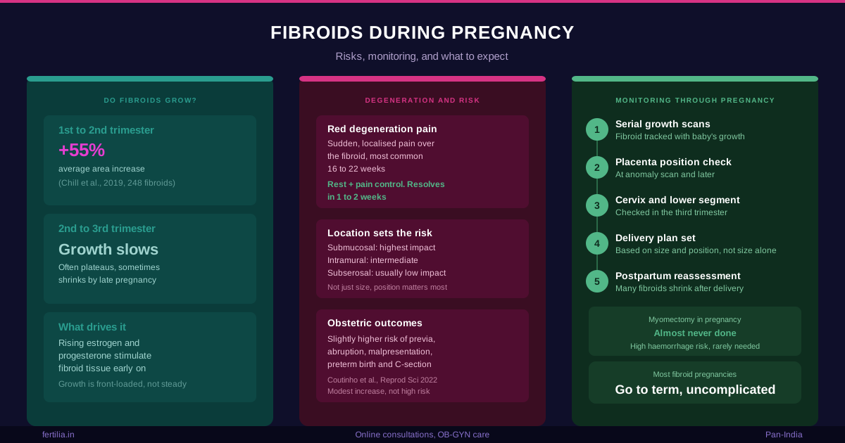
A separate cohort study out of Israel, tracking 248 fibroids across serial scans, put numbers to that pattern: fibroid area increased by roughly 55% on average between the first and second trimester, with a smaller and statistically non-significant increase between the second and third. Across the whole pregnancy, fibroids nearly doubled in area on average by the third trimester compared to the first, but the growth was front-loaded, not steady (Chill et al., BMC Women’s Health, 2019).
What drives this is estrogen and progesterone, both of which rise sharply in early pregnancy and are known to stimulate fibroid tissue, alongside changes in local blood supply as the uterus itself grows and remodels. Not every fibroid behaves the same way even within the same woman; if you have more than one, don’t be surprised if a follow-up scan shows one has grown while another hasn’t moved at all.
The plain-language version for your own report: fibroids most often grow early, tend to plateau or shrink later, and the overall change across a full pregnancy is usually modest, not dramatic. A fibroid that was 3 cm at your dating scan being 4 cm at 20 weeks is a normal, expected finding, not a red flag on its own.
Red degeneration: the pain that sends women to the emergency room
This is the complication that catches women completely off guard, usually because nobody warned them it was coming. Somewhere in the second trimester, most often between 16 and 22 weeks, a fibroid can outgrow its own blood supply. The tissue inside starts to break down, and the result is acute, often severe, localised pain directly over the fibroid, sometimes with mild fever and tenderness when the area is pressed.
This is called red degeneration, sometimes carneous degeneration, and it is the single most common reason a pregnant woman with a known fibroid ends up being scanned urgently or admitted for pain control. It is frightening when it happens. It is not, in itself, dangerous to you or your baby.
What it typically looks like:
- Sudden, sharp, well-localised pain directly over a known fibroid, not diffuse cramping
- Low-grade fever in some cases
- Tenderness when the area is pressed on examination
- Onset most commonly in the second trimester, when fibroid growth is at its fastest
How it’s managed: rest, paracetamol, and time. Red degeneration is a self-limiting event; the pain typically peaks over the first few days and resolves within one to two weeks as the tissue settles, even though it can be genuinely miserable while it lasts. Anti-inflammatory painkillers are generally avoided in pregnancy depending on gestational age, which is exactly why this is managed with your obstetric team rather than at home, so the right pain control for your stage of pregnancy gets used. Surgery during this event is almost never needed and, as covered below, is actively avoided unless there is no other option.
If you develop sudden, severe, localised abdominal pain in pregnancy, whether or not you know you have a fibroid, get it evaluated rather than waiting it out. The reassurance here is about what red degeneration usually turns out to be once assessed, not a reason to skip the assessment itself.
Location matters more than size
A 3 cm fibroid in one position can be a complete non-issue. A 3 cm fibroid in another position can shape your entire delivery plan. This is the single most important thing to understand from your scan report, and it’s why “how big is it” is often the wrong first question.
Submucosal fibroids sit closest to or bulge into the uterine cavity, the same space your baby is growing in. These carry the highest risk of affecting the pregnancy, because their position can interfere with placental attachment and growth space.
Intramural fibroids sit within the muscular wall of the uterus. Depending on size and how much they distort the cavity, these carry an intermediate risk, and many cause no issues at all.
Subserosal fibroids grow outward, on the outer surface of the uterus. These are generally the least likely to interfere with the pregnancy itself, even when they reach a significant size, because they don’t compete for space inside the cavity.
If your report also uses the FIGO type system (0 through 8, part of the FIGO PALM-COEIN classification for abnormal uterine bleeding; Munro MG et al., International Journal of Gynecology & Obstetrics, 2011; PMID 21345435) alongside these three terms, our guide to reading a fibroid ultrasound report, written by our consultant radiologist Dr. Rajashree NS, breaks down exactly what those numbers mean and why the classification changes management more than the size in centimetres does.
A large fibroid low in the uterus, particularly one sitting in the lower segment near the cervix, deserves specific mention. If it’s large enough and positioned in a way that could obstruct the birth canal, this is one of the more common reasons a planned caesarean section gets recommended for a woman who might otherwise have delivered vaginally. This is always an individual assessment based on your own scans as pregnancy progresses, not an automatic rule for every fibroid near the cervix.
What the evidence shows about obstetric risk
Here is where it’s worth being precise, because online searches on this topic swing between two unhelpful extremes: dismissing fibroids as irrelevant, or treating every fibroid pregnancy as high-risk. Neither is accurate.
A 2022 narrative review by Coutinho and colleagues, summarising the available cohort and case-control evidence, lays out where the risk actually sits: the presence of fibroids is associated with a slightly increased risk of placenta previa, placental abruption, and fetal malpresentation, largely from the mechanical effect of multiple or large fibroids taking up space in the uterus. Caesarean section and preterm birth rates are also probably somewhat higher in fibroid pregnancies. The review is explicit that these are modest, statistically demonstrated increases in risk, not high absolute risks, and that a causal relationship between fibroids and pregnancy loss specifically remains uncertain outside of women with multiple submucosal lesions (Coutinho et al., Reproductive Sciences, 2022).
What this means in practice: if you have fibroids, your pregnancy is watched a little more closely than it would be otherwise, largely through the ultrasound monitoring described below, and the overwhelming majority of these pregnancies still go on to be normal, term, healthy deliveries. Fibroids shift the odds slightly; they don’t rewrite them. Our guide on placenta previa covers what that specific finding means if it comes up on one of your scans, and our guide on preterm labour signs covers what to watch for and when to call your doctor, useful background regardless of whether fibroids are part of your picture.
Fibroids are common enough in pregnancy, reported in anywhere from roughly 1 in 10 to as few as 1 in 1,000 pregnancies depending on how they’re detected and counted, that most obstetric units in India, including the teams we work alongside at Fertilia, have a standard approach to monitoring them rather than treating each case as unusual (Cooper & Okolo, Obstetrical & Gynecological Survey, 2005).
How your doctor monitors a fibroid through pregnancy
If a fibroid is found on your dating scan, or you already knew about one going in, expect it to become a routine part of your antenatal visits rather than a separate concern:
- Serial growth scans, tracking the fibroid’s size alongside your baby’s growth at the visits where growth scans are already scheduled, not extra appointments purely for the fibroid
- Position relative to the placenta, checked at your anomaly scan and again later, since this relationship (not just the fibroid’s size alone) drives the placenta previa and abruption risk discussed above
- Position relative to the cervix and lower segment, particularly in the third trimester, to plan mode of delivery
- Symptom check-ins at each visit, specifically asking about the kind of sudden, localised pain that would suggest red degeneration
None of this changes how many antenatal visits you have or adds separate procedures. It’s the same monitoring schedule, with your fibroid factored into what’s being looked for at each scan.
If you’d like to talk through what your specific scan findings mean for your pregnancy, you can reach out to Dr. Suganya Venkat for a video consultation at Fertilia. Fertilia works with women across India, entirely online, through video and phone consultations.
Why myomectomy during pregnancy is almost never done
If you’ve read about fibroid surgery and are wondering whether it makes sense to just remove the fibroid now, while you’re already under medical care, the answer for the overwhelming majority of pregnancies is no.
The uterus during pregnancy has a dramatically increased blood supply, which is exactly what makes myomectomy in this setting so high-risk: the haemorrhage risk is substantial, entering the uterine cavity risks the membranes and the pregnancy itself, and the fibroid, as covered above, is likely to change size on its own as pregnancy progresses anyway. Myomectomy during pregnancy is reserved for the rare, specific circumstance where a fibroid is causing a complication, most often uncontrollable pain or torsion of a pedunculated fibroid, that cannot be managed conservatively by any other means, and even then it’s a decision made jointly between obstetrician and, often, a surgical team, weighing risk against risk. This is genuinely an exceptional scenario, not a routine option to bring up at a checkup.
If surgery for your fibroids does turn out to be the right path, it is planned for before conception or after delivery, when it can be done safely and thoroughly. Our guide to fibroid surgery (myomectomy) cost and recovery in India covers what that looks like when the timing is right, and our guide on conceiving naturally with fibroids covers the pre-pregnancy version of this same question, whether treatment is worth pursuing before you try to conceive at all.
After delivery: what happens to the fibroid
This is often the most reassuring part of the whole conversation, and it’s worth knowing before you’re in the thick of a fibroid pregnancy rather than after. Many fibroids reduce in size in the months following delivery, as the pregnancy hormones that stimulated growth fall away and the uterus itself returns to its pre-pregnancy size. Breastfeeding, through its hormonal effects, is thought to accelerate this regression in many women.
This doesn’t mean every fibroid disappears, and some women do go on to consider treatment postpartum if the fibroid remains symptomatic or is relevant to plans for a future pregnancy. But it does mean the fibroid found on your dating scan is not necessarily the fibroid you’ll be dealing with a year from now, and it’s genuinely reasonable to defer that decision until after you’ve had time to see what your body does on its own.
What you can do this week
- If a fibroid was found on your dating or anomaly scan, ask your doctor specifically about its location relative to the placenta and the cervix, not just its size
- If you develop sudden, localised, severe pain in pregnancy, get it evaluated promptly rather than assuming it’s normal pregnancy discomfort or waiting to see if it settles
- Keep your scheduled growth scans even if you feel completely well; this is how position and growth changes relevant to your delivery plan are tracked
- Don’t assume surgery is the next step. For the vast majority of fibroid pregnancies, watching and monitoring is the entire management plan
- If you’re planning your next pregnancy and already know you have fibroids, this is a reasonable time to discuss with your doctor whether pre-pregnancy treatment makes sense for your specific fibroid, rather than deciding mid-pregnancy
Frequently Asked Questions
Do all fibroids grow during pregnancy? Most fibroids grow to some degree in the first trimester, when estrogen and progesterone rise sharply. Growth typically slows in the second trimester and can plateau or even reverse by the third. Not every fibroid grows at the same rate, and some barely change at all across a full pregnancy.
Is fibroid pain in pregnancy dangerous? The most common cause of significant fibroid pain in pregnancy is red (carneous) degeneration, which is uncomfortable but not dangerous to you or your baby. It’s managed with rest and appropriate pain control and typically resolves within one to two weeks. Any sudden, severe abdominal pain in pregnancy should still be evaluated promptly to confirm that’s what it actually is.
Can a fibroid cause a miscarriage? A direct causal link between fibroids and pregnancy loss is not firmly established in the general population, according to the current evidence. The exception is women with multiple submucosal fibroids, where the risk does appear meaningfully higher. Most fibroid pregnancies do not end in miscarriage.
Will I need a C-section because of my fibroids? Not automatically. Caesarean rates are somewhat higher overall in fibroid pregnancies, but the decision depends on your specific fibroid’s size, position and relationship to the cervix and lower segment, assessed on your third-trimester scans, not on having a fibroid diagnosis at all.
Should my fibroid be removed during my C-section? This is generally avoided. Removing a fibroid at the same time as a caesarean significantly increases bleeding risk and operating time. Most obstetric teams leave fibroids untouched during delivery and revisit treatment afterward, if it’s still needed once the uterus has returned to its normal size.
Can I still have a vaginal delivery with fibroids? Yes, in most cases. The majority of women with fibroids in pregnancy deliver vaginally without complication. Whether a caesarean is advised comes down to your specific fibroid’s size and position, particularly whether it’s low enough to obstruct the birth canal, not to the presence of a fibroid on its own.
Do fibroids shrink after delivery? Many do, as pregnancy hormone levels fall and the uterus returns to its pre-pregnancy size, and breastfeeding may speed this along. This is one reason many women are advised to wait and reassess after delivery, rather than planning fibroid treatment during the pregnancy itself.
If you’ve found a fibroid on your pregnancy scan and want to understand what it means for your specific situation, whether that’s monitoring through the rest of your pregnancy or planning ahead for delivery, you can reach out to Dr. Suganya Venkat for a video consultation at Fertilia. Fertilia works with women across India, entirely online.