Two miscarriages in twelve months. The second one came with a referral for tests that had not been run before, including a 3D scan of the uterus. The report came back with a phrase many women encounter for the first time that same week: septate uterus.
If this is where you are, here is what that finding means, how it is diagnosed properly, what it does to a pregnancy when it is left in place, and what surgery can reasonably be expected to change.
What Is a Septate Uterus?
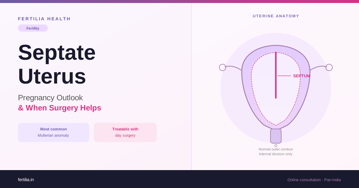
The uterus forms during fetal development when two channels called the Müllerian ducts fuse together. After they join, the wall of tissue between them normally dissolves, leaving a single, open cavity. When this reabsorption does not happen completely, a band of fibromuscular tissue remains and divides the cavity from the inside. That band is a uterine septum, and the condition is a septate uterus.
A review by Saravelos and colleagues, published in Human Reproduction Update in 2008, found that septate uterus was the most commonly identified uterine anomaly among women being investigated for reproductive failure. It sits at the top of a list that also includes arcuate, bicornuate, unicornuate, and didelphys uteri, each of which forms differently and is managed differently.
The septum itself varies in length. Some septa are short, projecting only slightly into the top of the cavity. Others are longer, reaching partway down, or occasionally extending all the way to the cervix, which is called a complete septum. Length matters clinically because a longer septum reduces the portion of the cavity available for safe implantation.
Why the distinction from a bicornuate uterus matters
Septate and bicornuate uteri are commonly confused, and the distinction has practical consequences. In a septate uterus, the outer shape of the uterus is entirely normal. The division is internal, inside the cavity, and the outer uterine wall forms a single dome. In a bicornuate uterus, the outer wall of the uterus itself is divided into two horns, producing a heart-shaped external contour that is visible on imaging.
This matters because the two conditions require different approaches. A septate uterus responds to a minimally invasive hysteroscopic procedure where the septum is removed through the cervix, without any external incisions. A bicornuate uterus requires a different and more complex surgical approach, which is not routinely offered for fertility purposes. Treating a bicornuate uterus as if it were a septate one, or vice versa, produces poor results, which is why the imaging step is not a formality.
An arcuate uterus, at the other end of the spectrum, is a mild indentation of the fundal wall that is generally considered a normal variant. It is not associated with the same rate of pregnancy loss and surgery is not routinely offered.
How a Septate Uterus Is Diagnosed
A standard 2D transvaginal scan or hysterosalpingogram (HSG) can suggest that the uterine cavity is divided, but neither test can reliably tell the difference between septate and bicornuate. Both conditions produce a similar appearance of a two-part cavity on these investigations, but the external uterine shape, which is the defining distinction, is not adequately assessed by either.
3D transvaginal ultrasound is the preferred first-line investigation for classifying uterine anomalies. It shows the inside of the uterine cavity and the outer contour of the uterus simultaneously, and a gynaecologist experienced in interpreting 3D scans can use it to make the septate-versus-bicornuate distinction with confidence in most cases. If you would like to understand what a transvaginal scan involves and what it can show, the post on Transvaginal Scan (TVS): What It Shows and Why It Is Done is a useful starting point.
MRI provides excellent soft tissue detail and is the investigation of choice when the 3D scan picture is not clear, when the uterine anatomy is particularly complex, or when additional information about the uterine wall is needed before a surgical decision. It is more expensive and less widely available at the community level in India, but for a decision that involves hysteroscopic surgery, the additional certainty it provides is worth the cost.
Saline infusion sonography (SIS) uses a small volume of saline instilled into the uterine cavity before scanning, which outlines the cavity and improves the visibility of any internal septum. It can be combined with 3D imaging for more precise characterisation of the anatomy.
Diagnostic hysteroscopy with laparoscopy was historically the gold standard because it allowed direct visualisation of the cavity from the inside (hysteroscopy) while checking the outer shape of the uterus simultaneously (laparoscopy). This remains the most definitive investigation when 3D imaging is equivocal, but for straightforward cases where the 3D scan gives a clear picture, this more invasive step is not always required.
How a Septum Affects Pregnancy
The core problem is the tissue itself. Unlike normal uterine wall, which has a rich blood supply and responds adaptively to the hormonal environment of early pregnancy, a septum is composed of fibromuscular tissue with significantly reduced vascularity. When an embryo implants on or near the septum, it cannot receive the blood supply needed to develop a properly functioning placenta. This is the primary mechanism behind the elevated miscarriage rate associated with septate uterus.
Most pregnancy losses attributable to a septum occur in the first trimester, typically between six and twelve weeks, when placentation is establishing itself. The pregnancy may begin with normal hormone levels and even a heartbeat on an early scan, and then fail to progress because the implantation site cannot sustain it.
Some women with a septate uterus do conceive and carry pregnancies without ever being diagnosed. In these situations, the embryo has implanted in an area of the cavity with normal blood supply, away from the septum. These women may discover the condition only when a scan is done for an unrelated reason, or when a pregnancy loss eventually prompts investigation.
A longer septum, because it occupies more of the cavity, increases the probability that any given implantation will land on or near it. This is not a certainty in either direction, but it shapes the clinical discussion about how urgently surgery should be considered.
Beyond miscarriage risk, a septum can also contribute to preterm birth in later pregnancy, abnormal placentation, and an increased likelihood of an abnormal fetal position near term. The divided cavity gives the baby less room to settle into the standard head-down position, which is why higher rates of breech and transverse lie are seen in women with untreated septate uterus.
If you have been diagnosed with a septate uterus and want to understand what it means for your pregnancy chances, you can speak with Dr. Suganya over a video consultation.
Who Benefits from Surgery, and Who Can Wait?
Not every woman with a septate uterus is in the same situation, and surgery is not automatically indicated on diagnosis alone. The decision depends on the clinical picture as a whole.
Surgery is typically recommended in these situations:
Recurrent miscarriage (two or more pregnancy losses) where other causes have been investigated and the septum is the most likely remaining contributor. The post on Recurrent Miscarriage: Tests After Two Losses covers what a thorough investigation should include before any structural cause is called definitive. Addressing the anatomy is more meaningful when the surrounding workup is clear.
Recurrent implantation failure in IVF cycles, where a normal-looking embryo is transferred but fails to implant or sustain, and the uterine environment is suspected as the factor. Removing a septum before a subsequent cycle has been associated with improved outcomes in this group. The post on Recurrent Implantation Failure: Causes and the ERA Test explains what the full workup in this situation looks like.
A substantial septum found incidentally in a woman preparing for a first pregnancy attempt, where surgical correction before trying makes more sense than waiting to see if a loss occurs first, particularly if the septum is long or if there are other factors that make natural conception difficult.
Surgery may not be the immediate priority when:
The septum is short or mild and the woman has not yet attempted pregnancy. Some gynaecologists take a cautious approach in this situation, particularly in younger women with no other fertility-limiting factors, where a trial of natural conception is a reasonable starting point. If losses follow, surgery is then indicated.
Other fertility-affecting factors are present, including a significantly reduced ovarian reserve, blocked fallopian tubes, or a substantial male factor issue. In these cases, attending to the other limiting factors alongside or before surgery produces a more complete picture.
The conversation about surgery is one to have with your gynaecologist, with the imaging and investigation results in front of you. A septum does not carry a one-size-fits-all recommendation.
What Hysteroscopic Septum Resection Involves
Hysteroscopic septum resection, sometimes called hysteroscopic metroplasty, is a day procedure performed under anaesthesia, typically general or spinal. A thin telescope-like instrument (the hysteroscope) is passed through the cervix into the uterine cavity, without any external incisions or cuts to the skin. The septum is divided using a fine cutting instrument, an electrical loop, or laser energy passed through the hysteroscope.
The procedure generally takes between 30 and 60 minutes depending on the length of the septum. Most women are discharged the same day. The minimally invasive nature of the approach means that the recovery time is substantially shorter than for older open surgical techniques.
Procedure-related risks include a rare risk of perforation of the uterine wall, fluid absorption into the circulation during the procedure (managed with careful fluid monitoring), and a small possibility of intrauterine adhesions forming as the tissue heals. These risks are not eliminated but are uncommon in experienced hands.
A follow-up scan or office hysteroscopy is usually arranged at six to eight weeks to confirm that the resection is complete and that the cavity has healed without adhesions. If the initial resection was incomplete, a second procedure is typically offered at this review, before clearing the patient to try to conceive. For context on procedure costs at Indian hospitals, the post on Hysteroscopy Cost in India 2026 covers what to expect at different types of facilities.
After Surgery: Pregnancy Outlook
The evidence on pregnancy outcomes after hysteroscopic septum resection comes largely from observational studies and case series. A smaller number of prospective studies exist, and randomised controlled trials are few, partly because the ethical challenges of allocating women with recurrent loss to a non-surgical arm are considerable.
What the published literature consistently shows is that among women with a septate uterus and a history of recurrent pregnancy loss, live birth rates after resection are meaningfully better than the pre-surgical baseline. The improvement in outcomes is most pronounced in women with a clear loss history that points to the septum as the contributing factor, and in whom the resection has been performed completely.
Dr. Suganya Venkat, an OB-GYN with over 15 years of practice managing uterine structural findings, takes a straightforward view: a septate uterus with a recurrent-loss history is one of the more satisfying problems to address precisely because the intervention is well-defined and reproducible. The anatomy changes in a way that can be confirmed on follow-up imaging, and for most women, the pregnancy outlook is genuinely better afterwards.
Waiting before trying to conceive
Most gynaecologists recommend waiting two to three months after hysteroscopic septum resection before trying to conceive. This waiting period allows the uterine lining to regenerate fully over the area where the septum was removed. Two full menstrual cycles is a common minimum; some clinicians recommend three cycles after a more extensive resection. Beginning attempts before the cavity has healed risks implantation into tissue that is still in the process of recovering.
After the waiting period, the natural conception process can begin as normal, or assisted conception protocols can be planned around the confirmed-healed uterus. Having a confirmed-healed cavity on imaging before starting an IVF cycle, if that is the next step, removes one variable from what is already a complex process.
If this applies to you, or if you are mapping out what comes next after a diagnosis or a loss, a clear consultation is the most useful first step. The Fertility Program outlines how we support women through exactly these decisions, from investigation through to conception.
Frequently Asked Questions
Do all women with a septate uterus need surgery?
Not necessarily. Some women with a septate uterus conceive and carry pregnancies without knowing the condition was present. Surgery is recommended when there is a history of recurrent pregnancy loss, repeated IVF failure where the uterus is the suspected factor, or when imaging shows a substantial septum before any pregnancy attempts and the decision is to optimise anatomy first. A short septum in a woman with no reproductive history yet is often watched rather than treated immediately.
Can I conceive naturally with a septate uterus without having surgery first?
Yes, it is possible. A septum does not block the fallopian tubes or prevent ovulation. The problem it creates is at the level of implantation quality: if the embryo settles on the septum, the blood supply there is inadequate for the placenta to develop properly. If the embryo implants away from the septum, in an area of the cavity with normal blood supply, the pregnancy may proceed normally. Whether to attempt natural conception before surgery depends on the length of the septum, your reproductive history, and your age.
How long after septum resection before I can try to conceive?
Most gynaecologists recommend two to three months, allowing time for the uterine lining to regenerate over the area where the septum was removed. A follow-up scan or hysteroscopy is usually done at around six to eight weeks to confirm the cavity has healed fully before you are cleared to try.
What is the difference between a septate and a bicornuate uterus?
In a septate uterus, the external shape of the uterus is normal and the division is entirely inside the cavity. In a bicornuate uterus, the outer uterine wall itself is split into two horns, giving it a visible heart-shaped external contour. The surgical treatment that works for a septate uterus (hysteroscopic resection) is not the right approach for a bicornuate uterus, which is why accurate diagnosis with 3D ultrasound or MRI is necessary before any surgical decision.
Can a septate uterus be seen on a routine 2D scan or HSG?
A 2D transvaginal scan or HSG can suggest a divided cavity, but neither test reliably distinguishes septate from bicornuate. A 3D transvaginal ultrasound, which shows both the inner cavity and the outer uterine contour at the same time, is the preferred tool. Where the 3D picture is unclear, MRI provides the additional detail needed.
Will I need a caesarean section because of a septate uterus?
Not automatically. After a successful septum resection and a healed cavity, most women can attempt vaginal delivery. A septate uterus does carry a higher rate of abnormal fetal position near term (because the divided cavity reduces the space for the baby to settle head-down), but delivery mode is decided on the clinical picture at the time. A corrected septate uterus does not mean caesarean by default.
If my septum resection was incomplete, what happens next?
A follow-up hysteroscopy or scan at six to eight weeks will usually show whether the resection was complete. If the septum is still partially present, a second procedure is typically offered before you start trying to conceive. Two-stage resections are not uncommon for longer septa, and the follow-up step exists precisely to catch and address incomplete resections before they affect a pregnancy.
A septate uterus is one of the more clearly defined and treatable causes of recurrent pregnancy loss. The anatomy can be confirmed on imaging, the intervention is minimally invasive, and the change it produces in the uterine cavity is one that can be seen on a follow-up scan. For many women, the period after resection is also the point at which the pregnancy that finally works out begins.
If you have received this diagnosis and want to work through what it means for your situation, you can speak with Dr. Suganya over a video consultation, from anywhere in India.