A woman messages a photo of her pelvic ultrasound report. Under “impression,” it reads: “Bilateral polycystic ovaries noted.” Her cycles are regular. Her blood tests are normal. But she has read online that polycystic ovaries mean PCOS, and she is now convinced she has a condition nobody has actually diagnosed her with.
I am Dr. Rajashree NS, a consultant radiologist, and I write reports with this exact line most weeks. What the ultrasound shows and what the diagnosis is are two different questions, and the report answers only the first one. This guide explains what a radiologist is actually describing when a report says “polycystic ovaries,” how that finding fits into the Rotterdam Criteria that gynaecologists use to diagnose PCOS, and why the scan by itself, in either direction, never settles the question.
What this post covers:
- What “polycystic ovaries” or “polycystic ovarian morphology” means on a report
- How radiologists count follicles and measure ovarian volume, and why the threshold changed in 2023
- The Rotterdam Criteria: the three features and why you need two of three
- Why some women have polycystic-appearing ovaries with no PCOS, and why some women with PCOS have normal-appearing ovaries
- What to do with your report once you have it
What “Polycystic Ovaries” Means on a Report
The word “polycystic” is one of the most misleading terms in gynaecology, because it does not describe cysts in the way most women picture them. There is no fluid-filled mass, no growth, nothing that needs removing.
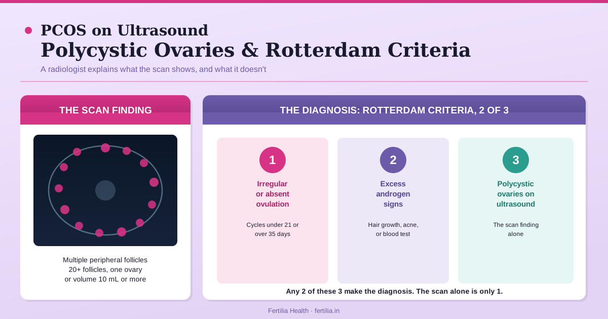
What a transvaginal ultrasound is actually measuring is the number and arrangement of small antral follicles, the fluid-filled sacs that hold immature eggs at their resting stage. In a polycystic-appearing ovary, these follicles are unusually numerous, often described as arranged around the edge of the ovary like a string of pearls, sometimes written on a report as the “necklace sign.” The ovary itself may also measure larger than typical.
A report describing this finding might read: “Both ovaries show increased stromal volume with multiple small peripheral follicles, right ovary measuring 11 cc, left ovary measuring 10.5 cc. Findings suggestive of polycystic ovarian morphology.” That sentence is a description of appearance, not a diagnosis. The report is telling the referring doctor what the ovary looks like on this scan, on this day. It is not telling you whether you have PCOS.
How the Count Is Done, and Why the Threshold Changed
For years, the working ultrasound threshold for polycystic ovarian morphology (PCOM) was more than 12 follicles in one ovary, or an ovarian volume of 10 mL or more. This came from the Rotterdam consensus criteria published in 2004, and it is still the number that appears in a lot of older reference material, including some patient-facing explainers online.
That threshold has since moved. The 2023 International Evidence-based Guideline for the Assessment and Management of Polycystic Ovary Syndrome (Teede HJ et al., Fertility and Sterility, 2023; PMID 37589624), developed by an international panel and endorsed across major reproductive medicine societies, raised the follicle-count threshold to 20 or more follicles in at least one ovary when a modern high-resolution transvaginal probe is used and the whole ovary can be counted clearly. Ovarian volume of 10 mL or more remains the fallback marker, used when the equipment is older or the full follicle count cannot be reliably done, including whenever a transabdominal rather than transvaginal probe is used.
Why the number moved up rather than down: newer ultrasound machines resolve smaller follicles than the machines available in 2004 did. Counting more follicles with better equipment was inflating how many ovaries got labelled “polycystic” using the old cutoff, so the threshold was raised to match what modern machines can actually see. If your report or your doctor’s explanation references the older “>12 follicles” figure, that is not wrong exactly, it reflects the criteria in place when a lot of currently practising radiologists trained. Ask which threshold and which probe your centre used if the number seems borderline.
Two details worth checking on your own report. First, whether the scan was transvaginal or transabdominal, since the reliable threshold differs between the two. Second, if you are counting follicles yourself from a report that lists them individually, the count that matters is the higher of your two ovaries, not the total added together.
The Rotterdam Criteria: Why the Scan Is One of Three
Polycystic ovarian morphology on ultrasound is one of three features used to diagnose PCOS under the Rotterdam Criteria (Rotterdam ESHRE/ASRM-Sponsored PCOS Consensus Workshop Group, Fertility and Sterility, 2004; PMID 14711538), refined further by the Androgen Excess and PCOS Society task force (Azziz R et al., Fertility and Sterility, 2009; PMID 18950759). A PCOS diagnosis requires at least two of the following three:
1. Ovulatory dysfunction. Irregular periods (cycles shorter than 21 days or longer than 35 days), infrequent periods, or absent periods, reflecting that ovulation is not happening consistently.
2. Clinical or biochemical hyperandrogenism. Signs of higher androgen levels than typical, such as excess facial or body hair growth, jawline acne, or hair thinning at the scalp (clinical), or an elevated testosterone or free androgen index on a blood test (biochemical).
3. Polycystic ovarian morphology on ultrasound. The follicle count or ovarian volume finding described above, in at least one ovary.
A woman needs two of these three. Polycystic-appearing ovaries alone, without irregular cycles and without any androgen excess sign, is one criterion out of three, and on its own it is not a PCOS diagnosis. This is the single most common misunderstanding I see attached to a scan report, and it is worth restating plainly: a scan finding is not a diagnosis by itself, under any of the three Rotterdam criteria taken alone.
The 2023 guideline (Teede et al., PMID 37589624) also clarified that if a woman already has both irregular cycles and confirmed hyperandrogenism, an ultrasound is not required at all to make the diagnosis. Some women are diagnosed with PCOS on history and blood tests alone, with a scan never entering the picture, or a scan done later that happens to look entirely normal.
Polycystic Ovaries Without PCOS
Polycystic-appearing ovaries are a genuinely common incidental finding. Research going back to the early ultrasound era, when Clayton et al. (Clinical Endocrinology, 1992; PMID 1395063) scanned a group of women with no known reproductive complaints, found polycystic ovarian morphology in a substantial proportion who had entirely regular cycles and no hormonal abnormality. A polycystic-appearing ovary, in other words, is not automatically a sign that anything is wrong.
If your ultrasound found polycystic-appearing ovaries and your periods come reasonably on schedule, and you have no facial hair, acne, or abnormal androgen levels on a blood test, you do not meet the Rotterdam Criteria for PCOS on the strength of the scan alone. What you have is a normal anatomical variant that happens to look similar to what is seen in PCOS. Your OB-GYN may still note it in your file, since it means monitoring is sensible if your cycles change later, but it does not by itself require treatment.
PCOS Without Polycystic-Looking Ovaries
The reverse also happens, and it surprises women just as often. A woman can be clinically diagnosed with PCOS, meeting the ovulatory-dysfunction and hyperandrogenism criteria clearly, and still have an ultrasound that comes back describing normal-appearing ovaries. This is more common in what is sometimes called the “lean” or non-classic presentation, and it does not undo the diagnosis. Two of three criteria is the rule; if the other two are clearly present, a normal-looking scan does not rule PCOS out.
If you have been told you have PCOS based on your cycles and blood work, and a later scan looks unremarkable, that is not a contradiction. It reflects that morphology is only one of the three windows into the same underlying picture, and it does not always show up the same way on every scan, on every cycle.
If you are holding a report that mentions polycystic ovaries and are not sure what it means for you specifically, a video consultation puts your scan alongside your cycle history and, if needed, blood work, rather than reading the ultrasound line in isolation.
Dr. Suganya Venkat sees women for this exact question through Fertilia’s online video consultations, Rs. 399, pan-India. You can share your report directly in the conversation.
What the Rest of Your Report Might Also Show
A pelvic ultrasound done for PCOS workup often includes a few other lines worth understanding, since they sometimes cause separate confusion.
Endometrial thickness. With infrequent or absent periods, the uterine lining can build up over a longer stretch of time than usual. Your report may note this as a specific millimetre measurement. This is a monitoring detail your OB-GYN factors into whether and how to bring on a period, not an emergency finding on its own.
Ovarian volume individually. Some reports list the two ovaries separately rather than giving one combined impression. It is common for one ovary to look more classically polycystic than the other. Only one ovary meeting the threshold is enough to satisfy that criterion.
“No dominant follicle seen.” On a scan done later in the expected cycle, this notes that ovulation has probably not yet occurred by that point. On its own, this is consistent with the irregular-ovulation pattern common in PCOS, but a single scan on a single day is a snapshot, not a full ovulation assessment. For how ovulation itself is confirmed with blood work rather than a single scan, our guide on antral follicle count and reading your ultrasound covers the related but distinct question of counting follicles for fertility-reserve purposes rather than for PCOS diagnosis.
Why This Distinction Matters for What Happens Next
Getting this right changes what kind of conversation you have with your doctor. A woman with polycystic-appearing ovaries but no other Rotterdam criterion generally needs reassurance and routine monitoring, not a PCOS treatment plan. A woman who meets two or three criteria, including morphology, benefits from a fuller conversation about how PCOS is actually managed, insulin sensitivity, cycle regulation, and fertility planning if relevant, none of which follows from the ultrasound line by itself.
If your ultrasound is one part of a picture that also includes irregular periods, acne, or hair growth, our guide on PCOS: symptoms, root causes, and treatment walks through the fuller diagnostic and management picture. If your main question was about ovarian reserve rather than PCOS specifically, the AMH vs AFC guide explains the distinct reserve-testing purpose those same follicle counts can serve. And if heavier or irregular bleeding was what sent you for the scan in the first place, heavy periods after 35 separates PCOS from fibroids and adenomyosis as distinct causes that a single report can sometimes show together.
Frequently Asked Questions
Does “polycystic ovaries” on my ultrasound mean I have PCOS?
Not by itself. Polycystic ovarian morphology on ultrasound is one of three Rotterdam Criteria used to diagnose PCOS. A diagnosis needs at least two of the three: irregular ovulation, signs of excess androgens (clinical or on a blood test), and this ultrasound finding. If your cycles are regular and your androgen levels are normal, polycystic-appearing ovaries on a scan alone do not meet the threshold for a PCOS diagnosis.
What is the follicle count threshold for polycystic ovaries on ultrasound?
The current threshold, from the 2023 international PCOS guideline, is 20 or more follicles in at least one ovary on a modern high-resolution transvaginal scan, or an ovarian volume of 10 mL or more when a full follicle count is not possible. This replaced the older threshold of more than 12 follicles used since the 2004 Rotterdam criteria, updated because newer ultrasound machines resolve smaller follicles than older equipment could.
Can I have PCOS with a normal-looking ultrasound?
Yes. A woman with clearly irregular ovulation and confirmed hyperandrogenism, the other two Rotterdam criteria, can still be diagnosed with PCOS even if a scan shows normal-appearing ovaries. Morphology is one of three windows into the same condition, and it does not show up identically in every case or on every scan. The 2023 guideline also notes that ultrasound is not required at all if the other two criteria are already clearly met.
What does the “necklace sign” mean on a PCOS ultrasound?
The necklace sign describes multiple small follicles arranged around the outer edge of the ovary, resembling a string of pearls. It is a descriptive term for one visual pattern of polycystic ovarian morphology, not a separate diagnosis or a more severe finding than any other description of the same morphology.
Is polycystic ovarian morphology dangerous or does it need treatment?
Polycystic-appearing ovaries by themselves, in a woman with regular cycles and normal androgen levels, are a common incidental finding that generally does not require treatment. Treatment decisions follow from a full PCOS diagnosis, meeting at least two Rotterdam criteria together with your symptoms and goals, not from the ultrasound appearance in isolation.
How is a PCOS ultrasound different from a fertility reserve scan?
They can be the same scan read for two different purposes. A PCOS-diagnosis scan looks at follicle number and arrangement to assess morphology against the Rotterdam threshold. A fertility reserve scan (antral follicle count, done specifically on Day 2 to 5 of the cycle) uses a related follicle count to estimate ovarian reserve for fertility planning or IVF response. The counting method overlaps, but the clinical question and the timing requirement are different.
Should I get a repeat ultrasound if my report says polycystic ovaries?
Not urgently, and not on its own. If the finding was incidental and your cycles and androgen levels are normal, routine follow-up as part of your regular gynaecological care is generally sufficient. If you have irregular periods or symptoms of excess androgens alongside the scan finding, the next step is blood work and a fuller clinical assessment rather than simply repeating the same scan.
My report says polycystic ovaries but my periods are regular. Should I be worried?
Generally, no. Regular cycles mean you are very likely ovulating consistently, which is the criterion polycystic-appearing ovaries most commonly accompanies when PCOS is present. If your cycles are regular and you have no signs of excess androgens, this is most likely an incidental anatomical finding rather than PCOS, and it is a reasonable, common question to raise at your next routine visit rather than something that needs an urgent appointment.
A polycystic ovary finding on your scan report is information, not a verdict. What it means for you depends on the rest of your clinical picture, your cycles, your androgen levels, and your symptoms, read together rather than the ultrasound line on its own.
Message Fertilia on WhatsApp to go through your report and your full picture together, over a video consultation with Dr. Suganya Venkat, Rs. 399, wherever you are in India.
For the complete workup Fertilia uses to manage PCOS beyond a single scan, see the PCOS Symptom Reversal program.
Dr. Rajashree NS is a Consultant Radiologist with an MD in Radio-diagnosis from Sree Mookambika Institute of Medical Sciences. She interprets pelvic and fertility ultrasound reports as part of Fertilia’s team.