If you have just been told you have a molar pregnancy, or your scan report used the term “hydatidiform mole” and you are trying to understand what that means, you are probably feeling two things at once: grief for a pregnancy that will not continue, and confusion about a diagnosis that does not sound like the miscarriage you may have expected. Both reactions are entirely reasonable. A molar pregnancy is a different clinical event from a typical early pregnancy loss, with its own path forward: treatment followed by a period of monitoring. It is also, in the great majority of cases, very treatable, with excellent odds of a normal pregnancy afterward.
This guide explains what a molar pregnancy actually is, how it is diagnosed, what treatment involves, why the monitoring period afterward matters, and what your future fertility looks like once you are through it.
What this post covers:
- What a molar pregnancy is, and the difference between a complete and partial mole
- The signs that lead to diagnosis, and how a scan and hCG levels confirm it
- What treatment (evacuation) involves
- Why hCG monitoring afterward is essential, and what it involves
- The small risk of persistent disease, and how it is managed
- What your future pregnancy outlook looks like
What Is a Molar Pregnancy?
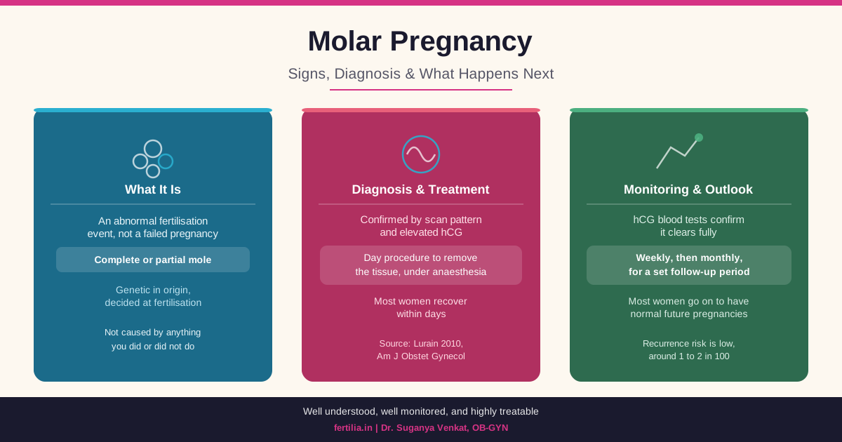
A molar pregnancy, medically called a hydatidiform mole, is not a typical pregnancy that has failed. It is a different event altogether: an error at fertilisation that causes the placental tissue (trophoblastic tissue) to grow abnormally instead of developing normally alongside an embryo. This abnormal growth, and how the body needs to be monitored afterward, is why molar pregnancy is managed differently from a standard miscarriage (Lurain JR, American Journal of Obstetrics and Gynecology, 2010, PMID 20728069).
There are two types, and the distinction matters for what you can expect:
Complete mole. This happens when an egg with no genetic material of its own is fertilised by one or two sperm. There is no embryo, no fetal tissue, no amniotic sac. All of the developing tissue is abnormal placental tissue. Because the egg contributed no genetic material, the resulting tissue’s chromosomes come entirely from the father’s side.
Partial mole. This happens when a normal egg is fertilised by two sperm, or by one sperm that duplicates its own genetic material, resulting in an abnormal extra set of chromosomes. Some fetal tissue does form in a partial mole, but it carries this chromosomal abnormality and is never capable of developing into a viable pregnancy.
In both cases, the underlying issue is genetic, decided at the moment of fertilisation. It has nothing to do with anything you did or did not do during the pregnancy, and in the overwhelming majority of cases, it does not reflect anything about your general fertility or the health of a future pregnancy.
Molar pregnancy is uncommon, and estimates of how uncommon it is vary by population, with somewhat higher reported rates across parts of Asia compared to Western countries, though good India-specific incidence data is limited. If you have been told you have one, it can feel like an isolated, frightening diagnosis. It is not common, but it is well understood, well studied, and has a clear, well-established management pathway that OB-GYN teams follow routinely.
Signs That Lead to Diagnosis
A molar pregnancy is usually picked up in one of two ways: symptoms that prompt an early scan, or a routine early-pregnancy scan that shows a characteristic pattern before any symptoms appear.
Vaginal bleeding in the first trimester is the most common presenting sign, and it is often the reason a woman comes in for a scan in the first place. It can range from light spotting to heavier bleeding, and it is sometimes accompanied by the passage of grape-like clusters of tissue, though this is a later and less common presentation than it once was, since most moles are now caught earlier on routine scanning.
A uterus that measures larger than expected for the dates, on clinical exam or on scan, is a classic sign of a complete mole in particular, caused by the rapid abnormal growth of the trophoblastic tissue.
Unusually severe nausea and vomiting, beyond typical morning sickness, can occur because hCG levels in a molar pregnancy are often disproportionately high for the gestational age, and hCG is part of what drives pregnancy nausea.
hCG levels that are markedly higher than expected for how many weeks pregnant you are is frequently the first clue, often picked up when a blood hCG test is done alongside an early scan.
Less commonly, especially with very high hCG levels, some women develop early signs of preeclampsia (high blood pressure with other features) unusually early in pregnancy, or symptoms of an overactive thyroid, since very high hCG can weakly stimulate the thyroid gland.
It is worth saying plainly: many women have no symptoms at all before their molar pregnancy is picked up on a routine first-trimester scan, which is increasingly how the diagnosis is made in India today, well before heavier bleeding or other signs would have appeared.
How the Diagnosis Is Confirmed
Ultrasound is the primary diagnostic tool. A complete mole has a distinctive appearance on scan, often described as a “snowstorm” pattern, a uterus filled with many small cystic spaces instead of a normal gestational sac and embryo. There is no fetal pole, no fetal heartbeat, and no amniotic fluid. A partial mole is sometimes less obvious on scan, since some fetal or placental tissue may be visible alongside the abnormal areas, which is part of why a partial mole is occasionally not suspected until after tissue is examined following an evacuation for what looked like a routine miscarriage.
Blood hCG levels support the diagnosis, particularly when they are markedly elevated relative to the gestational age. hCG alone does not confirm a molar pregnancy (many complications can raise or lower it), but a very high level combined with the ultrasound pattern is a strong indicator.
Histopathology after evacuation is the definitive confirmation. Whatever the scan suggests, the tissue removed during treatment is always sent for pathology examination, which confirms the diagnosis with certainty and distinguishes complete from partial mole. This step also matters for the monitoring plan that follows, since the two types carry slightly different follow-up considerations.
If your scan report mentions a “snowstorm pattern,” “vesicular pattern,” or “molar changes,” and you are waiting for a formal diagnosis, this combination of ultrasound findings, hCG level, and eventual pathology is the standard diagnostic sequence your care team is working through.
If you have just received this diagnosis and want to talk through what it means for you specifically, message me on WhatsApp and we can go through your scan and next steps together over a video consultation.
Treatment: What Evacuation Involves
The standard treatment for a molar pregnancy is suction curettage, a procedure very similar in principle to the surgical management used for a missed miscarriage, performed under anaesthesia to gently remove the abnormal tissue from the uterus. It typically takes around twenty to thirty minutes and is done as a day procedure at most hospitals in India, meaning most women go home the same day or the next.
Anti-D immunoglobulin is given after the procedure if you are Rh-negative, the same precaution used after any pregnancy loss in an Rh-negative woman, to prevent complications in a future pregnancy.
In the small number of cases where a molar pregnancy is diagnosed later, or where there are specific concerns, a hysterectomy may occasionally be considered, but this is reserved for particular circumstances, usually in women who have completed their family and have other clinical reasons to consider it. For the great majority of women, especially those who want to have children in future, suction curettage is the standard and preferred approach, and it preserves the uterus and future fertility.
You will likely feel physically better within days of the procedure, similar to recovery after a D&C for any other pregnancy loss. Light bleeding and cramping for a week or two afterward is normal.
Why hCG Monitoring Afterward Matters
This is the part of molar pregnancy care that differs most from a typical pregnancy loss, and it is worth understanding clearly rather than finding it confusing later.
After evacuation, your hCG level should fall steadily and become undetectable within a matter of weeks. Your doctor will arrange blood hCG tests at regular intervals, usually weekly at first, to confirm this downward trend. Once hCG becomes undetectable, monitoring continues monthly for a further period, in line with current international guidance on gestational trophoblastic disease follow-up (Ngan HYS et al., International Journal of Gynaecology and Obstetrics, 2018, PMID 30306586). Your own doctor will confirm the exact monitoring schedule and duration for your specific case, since it can vary slightly with the type of mole and how the numbers behave.
This monitoring is not optional, and it is not just a formality. In a small proportion of cases, a small amount of abnormal trophoblastic tissue persists after evacuation and continues producing hCG. This is called persistent gestational trophoblastic disease, and it is picked up precisely because you are being monitored, at a point when it is highly treatable, usually with medication alone. Skipping monitoring appointments is the main reason this would be missed, not the underlying condition itself being dangerous when caught on schedule.
Reliable contraception is required during the entire monitoring window. This is not a restriction on your life; it is a practical necessity for the monitoring to work. A new pregnancy during this period would itself raise hCG, and there would be no way to tell whether a rising number meant a new pregnancy or persistent disease. Your doctor will help you choose a contraceptive method that suits you for this window, most commonly a hormonal method or a barrier method, and this is a temporary measure tied specifically to the monitoring period, not a long-term restriction on your fertility.
The Small Risk of Persistent Disease, Explained Plainly
Reading about “gestational trophoblastic disease” or “persistent GTD” online can sound alarming, so it is worth being specific and calm about what this actually means. In a minority of women, most often after a complete mole, some abnormal tissue remains after evacuation and needs additional treatment, almost always chemotherapy, which for this specific condition has excellent, well-established cure rates. This is caught early precisely because of the hCG monitoring described above, which is the entire point of the follow-up schedule.
This is not something you need to watch for yourself day to day. It is something your hCG blood tests are watching for on your behalf. Your responsibility during this window is straightforward: attend the scheduled blood tests, use reliable contraception, and contact your doctor if you notice heavy bleeding, since these are the practical steps that let the monitoring system do its job.
Your Future Pregnancy Outlook
This is usually the question at the front of every woman’s mind once the immediate treatment is behind her, and the answer is genuinely reassuring. The overwhelming majority of women who have had a molar pregnancy go on to have completely normal future pregnancies, with no increased risk of miscarriage, birth defects, or other complications attributable to the mole itself (Joneborg U et al., International Journal of Gynecological Cancer, 2021, PMID 33649007). The recurrence risk (having another molar pregnancy in a future pregnancy) is low, on the order of one to two in a hundred, meaning the great majority of women never experience it again.
Once your monitoring period is complete and your doctor confirms it is safe to stop contraception, there is no medical reason to delay trying to conceive again if you feel ready. Many doctors, once monitoring has concluded, will suggest an early scan in your next pregnancy simply as a reassurance step, given your history, not because anything is expected to be different.
If a follow-up pregnancy comes and goes smoothly, most women find that this chapter, once frightening and confusing, becomes a distant part of their history rather than something that shapes the pregnancies that follow.
Frequently Asked Questions
What are the first signs of a molar pregnancy?
The most common early signs are first-trimester vaginal bleeding, a uterus that measures larger than expected for the dates, unusually severe nausea and vomiting, and an hCG level that is markedly higher than expected for the gestational age. Many molar pregnancies today are picked up on a routine early scan before any of these symptoms become noticeable.
How is a molar pregnancy diagnosed?
Diagnosis combines an ultrasound scan, which typically shows a characteristic pattern of many small cystic spaces instead of a normal pregnancy, with a blood hCG level that is often much higher than expected for the gestational age. The diagnosis is confirmed definitively when the tissue removed during treatment is examined under a microscope.
What is the difference between a complete and partial molar pregnancy?
A complete mole has no fetal tissue at all, only abnormal placental tissue, and occurs when an egg with no genetic material is fertilised. A partial mole has some fetal tissue, which carries an abnormal extra set of chromosomes and is never capable of surviving. Both are diagnosed and treated in largely the same way, though your doctor will tell you which type you had once pathology results are back, since it can slightly affect your monitoring plan.
How is a molar pregnancy treated?
The standard treatment is suction curettage, a day procedure performed under anaesthesia to remove the abnormal tissue from the uterus, similar to the procedure used for a missed miscarriage. Hysterectomy is reserved for specific circumstances and is not the standard approach, especially for women who want future pregnancies.
Why do I need hCG monitoring after treatment, and for how long?
hCG monitoring confirms that all the abnormal tissue has cleared and catches the small chance of any tissue persisting, at a point when it is highly treatable. It typically starts weekly and moves to monthly once levels are undetectable, continuing for a period your doctor will specify based on your individual case and mole type.
Can I get pregnant again after a molar pregnancy?
Yes. The great majority of women who have had a molar pregnancy go on to have completely normal future pregnancies. You will need to complete your hCG monitoring period and use reliable contraception until your doctor confirms it is safe to stop, but there is no reason to expect a future pregnancy to be affected once that window has closed.
Will a molar pregnancy happen again in my next pregnancy?
The recurrence risk is low, roughly one to two in a hundred, so the large majority of women who have had one molar pregnancy never have another. Because of this small increased risk, many doctors suggest an early reassurance scan in your next pregnancy, simply as routine precaution given your history.
Is a molar pregnancy the same as a miscarriage?
No. A miscarriage is the loss of a pregnancy that started developing normally. A molar pregnancy is a different event from the start: an abnormal fertilisation that never had the potential to develop into a viable pregnancy. The emotional experience of loss is real either way, and the grief deserves the same care, but the medical management and the follow-up monitoring afterward are specific to a molar pregnancy and different from a standard miscarriage.
A molar pregnancy diagnosis can feel disorienting because it is less familiar than a typical miscarriage, and because of the monitoring period that follows treatment. At Fertilia, Dr. Suganya Venkat has supported women through this exact diagnosis, from the moment of the scan through the monitoring window and into a future pregnancy when they are ready. If you have questions specific to your situation, message us on WhatsApp and we will go through it together, over a video consultation, at your own pace.
For related reading, see our guides to missed miscarriage, what your beta hCG numbers mean, coping after pregnancy loss, and tests to get after recurrent miscarriage. You can also download our Miscarriage Support Guide for a printable companion covering physical recovery and emotional healing.