You are sitting in the scan room at eleven or twelve weeks, watching the screen. The radiologist adjusts the image, moves the probe, types a number. On your report it will read something like “NT: 2.8 mm” or “NT: 3.6 mm.” The printout is handed over and the next patient is called. On the drive home, those few millimetres become the thing you cannot stop thinking about.
For more on this, read our guide on Growth Scan & Doppler in Pregnancy. The nuchal translucency measurement is one of the most important values in a first-trimester scan. It is also one of the most misread, partly because it appears as a single small number with no surrounding context. This guide explains exactly what that number measures, what a normal range looks like, what a raised value means, and what the steps are if further evaluation is recommended.
I am a radiologist. I write this report every working day. Here is what I would say to you if you were sitting across from me.
What this post covers:
- What the NT scan is and why the timing is precise
- How the measurement is taken, and why technique matters
- What normal NT ranges look like
- How NT combines with blood markers to give a risk assessment
- What a raised NT means, and what it does not mean
- The step-by-step pathway after a raised result
- Getting a good NT scan in India
What the NT Scan Is, and Why the Timing Is Exact
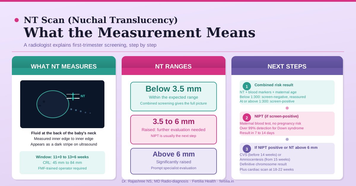
The NT scan is a first-trimester ultrasound done between 11 weeks and 0 days (11+0) and 13 weeks and 6 days (13+6) of pregnancy. This window is not chosen arbitrarily. It corresponds to the time when the baby’s crown-rump length (CRL) falls between 45 mm and 84 mm: large enough for the sonographer to place callipers precisely, small enough for the full anatomy to fit comfortably on screen in a midline view.
What the scan measures is the nuchal translucency: a fluid-filled space at the back of the baby’s neck, between the skin and the underlying subcutaneous tissue. On ultrasound, fluid transmits sound waves without scattering them, which means the space appears as a dark stripe at the nape of the neck. The sonographer places two calliper markers across this stripe and the software records the distance between them.
All fetuses carry some fluid in this space during the first trimester. As the lymphatic system matures, usually around 14 to 15 weeks, this fluid drains naturally. That is the other reason the scan cannot be done after 13+6 weeks: the translucency resolves, and the measurement is no longer valid.
How the Measurement Is Taken
This is worth understanding because the same fetus can produce a slightly different NT reading depending on technique, and knowing this helps you interpret your own result without alarm.
For a valid measurement, the sonographer needs:
- A midline sagittal view: a true side-profile of the fetus with the spine running horizontally across the screen, not rotated or tilted
- The fetal head in a neutral position: neither tucked toward the chest (hyperflexion, which decreases the measured NT) nor extended backward (hyperextension, which increases it)
- The image magnified so the fetus fills at least 75% of the screen: a small image produces an inaccurate calliper placement
- Callipers placed inner edge to inner edge at the widest point of the translucent space: not outer-to-outer, not at the skin surface
- At least three separate measurements taken; the best one, not the average, is recorded
The Fetal Medicine Foundation (FMF) developed and maintains the training and certification programme specifically for NT measurement. A sonographer who holds FMF certification has demonstrated technical competency in this exact technique, assessed against a reference dataset. Measurements from FMF-trained operators feed directly into the risk calculation software that produces the combined risk ratio on your report.
In India, most large diagnostic centres in metro cities (Chennai, Mumbai, Delhi, Bengaluru, Hyderabad) employ at least one FMF-trained or equivalently trained sonographer. In smaller cities, it is reasonable to ask before booking.
What the Numbers Mean: Normal NT Ranges
NT increases with gestational age. A measurement of 1.8 mm at 11 weeks is different from 1.8 mm at 13+6 weeks, because the baby and the fluid space grow over those three weeks. This means the NT value cannot be read in isolation without knowing the exact gestational age or CRL.
The standard clinical threshold used by most centres is NT below 3.5 mm. Below this value at any point in the 11+0 to 13+6 window is generally considered within the expected range.
The more precise way to express NT is as a multiple of the median (MoM): your measurement divided by the median expected for a fetus of exactly that CRL on that day. An NT of 1.5 MoM means the measurement is 1.5 times the expected median for that gestational age. This adjustment for gestation is the same method used for PAPP-A and free beta-hCG in the double marker blood test, and it is the value that enters the combined risk calculation software.
To make the numbers tangible: at a CRL of approximately 65 mm (around 12+2 weeks), the median NT is roughly 1.6 mm. An NT of 3.5 mm at that gestational age falls above the 99th centile for that CRL.
The landmark study by Snijders et al., published in The Lancet in 1998 (PMID 9717920), examined more than 100,000 pregnancies at the Fetal Medicine Foundation. It established that NT measurement combined with maternal age detected approximately 75% of trisomy 21 (Down syndrome) pregnancies, with a 5% false-positive rate. This study formed the evidence base for first-trimester NT screening as we still practice it today.
NT as Part of Combined First-Trimester Screening
The NT measurement is most useful when it is not standing alone. It is designed to be part of the combined first-trimester screening package: NT scan plus two blood markers, PAPP-A (pregnancy-associated plasma protein-A) and free beta-hCG, collected through a single maternal blood draw done at the same visit or within a few days.
In pregnancies affected by Down syndrome, the placenta tends to produce less PAPP-A than expected and more free beta-hCG than expected. The NT tends to be above the median. The laboratory software takes all three MoM values, adds the maternal age-related background risk, and computes a single combined risk ratio for trisomy 21, trisomy 18 (Edwards syndrome), and trisomy 13 (Patau syndrome).
A combined risk of 1:2,000 means 1 in 2,000 pregnancies with exactly this marker combination has the condition being screened for. A combined risk of 1:100 means 1 in 100. Most centres define screen-negative as a combined risk below 1:300 or 1:250 (the threshold varies slightly by laboratory). Screen-positive means the combined risk is at or above the threshold. Screen-positive is an instruction to look more closely, not a diagnosis.
For a complete explanation of the blood marker side of this test, including what PAPP-A and free beta-hCG values mean and what the risk ratio format looks like, read our double marker test guide.
If you have received a first-trimester screening result and are not sure what it means for your pregnancy, Dr. Suganya’s team can walk you through the result in the context of your full history. We work online, pan-India, by video call. Start a conversation on WhatsApp.
What a Raised NT Means, and What It Does Not
This is where I want to be careful and specific, because this is the part most patients find most difficult.
A raised NT is not a diagnosis. It is a screening finding. It tells you that this pregnancy has a higher statistical chance of a chromosomal or structural issue than a pregnancy with a lower NT. It does not tell you that the baby has a problem. Most pregnancies with mildly raised NT will be entirely unaffected.
The associations depend on the degree of elevation:
NT between 3.5 mm and 4.5 mm (mildly raised). The large majority of pregnancies in this range, particularly when the combined risk ratio remains low, go on to be completely normal. A closer look is appropriate and is the standard of care.
NT between 4.5 mm and 6 mm (moderately raised). A thorough evaluation is needed. Many pregnancies in this range also have normal chromosomes and no structural problems. The next step is NIPT and, depending on the combined risk, consultation with a fetal medicine specialist.
NT above 6 mm (significantly raised). This warrants evaluation without delay. Even here, a number of pregnancies will have a completely normal outcome. But the probability of a chromosomal or structural finding is meaningfully higher, and prompt evaluation matters.
The range of conditions associated with raised NT includes chromosomal differences (most commonly trisomy 21, trisomy 18, trisomy 13, and Turner syndrome), congenital heart defects, rarer genetic syndromes, and in some cases no identifiable cause at all. The heart is forming actively during the first trimester, and raised NT can reflect early differences in cardiac development that would be visible on a detailed cardiac scan later in pregnancy.
What Happens Next: The Step-by-Step Pathway
Step 1: The combined risk result. The NT, blood markers, and maternal age produce a single combined risk number. If the risk falls below the threshold, screen-negative, no further chromosomal screening is routinely recommended, though your doctor may arrange a follow-up scan or cardiac assessment depending on the degree of NT elevation.
Step 2: If screen-positive, NIPT is usually recommended first. NIPT (non-invasive prenatal testing), also called cell-free DNA screening or cfDNA testing, is a maternal blood test that can be done from 10 weeks onwards. It analyses fragments of fetal DNA circulating in the mother’s blood. For trisomy 21, NIPT has a detection rate above 99% with a false-positive rate below 0.1% (Gil MM et al., Ultrasound in Obstetrics and Gynecology, 2015, PMID 25639627). It carries no risk to the pregnancy. Results are typically available within 7 to 14 days from most accredited laboratories.
NIPT is a screening test. It screens for chromosomal differences but cannot diagnose them definitively. Diagnosing a chromosomal condition definitively requires testing a sample of the baby’s own cells.
Step 3: If NIPT is positive, inconclusive, or if NT is significantly raised (above 6 mm). Invasive testing is offered: chorionic villus sampling (CVS) before 14 weeks, or amniocentesis from 15 weeks onward. Both procedures take a small sample containing the baby’s cells for chromosome analysis. Both carry a small procedural risk of pregnancy loss, approximately 0.5 to 1%. This is why they are not the first step after a screen-positive result. They provide a definitive chromosome result where NIPT cannot.
Step 4: Cardiac assessment. For pregnancies with raised NT, regardless of the chromosome result, a detailed cardiac scan (fetal echocardiography) is usually arranged at around 18 to 22 weeks. The anatomy of the heart is best visualised at this gestation. This is in addition to, not instead of, the anomaly scan (TIFFA scan) that all pregnancies undergo at 18 to 22 weeks.
At each step, your obstetrician or fetal medicine specialist will explain the specific result and the options. The decisions about which tests to proceed with, and what to do with any result, belong to you.
Getting a Good NT Scan in India: Practical Points
Book within the right window. The NT scan must be done between 11+0 and 13+6 weeks of gestation. If your dates are uncertain (irregular cycles, unsure of last period), a dating scan at 7 to 9 weeks will give an accurate gestational age before you book the NT. Arriving for the NT scan at 14 weeks means the window has closed and the measurement will no longer be valid for combined risk calculation.
Ask about operator training. In large cities, FMF certification or equivalent training is standard at most reputable diagnostic chains. In smaller towns, asking this question before booking is a reasonable step.
The combined package gives more information than either test alone. The NT scan combined with PAPP-A and free beta-hCG blood markers gives a combined risk, which is far more informative than NT alone. Most hospitals and diagnostic chains offer this as a combined first-trimester screening package. For current pricing, the double marker test guide covers the cost ranges across private labs and hospitals in India.
If the baby was not in a cooperative position, a repeat scan may be needed. A good sonographer will not report an NT measurement from a technically inadequate image. If the report notes “baby not cooperative, repeat scan within 2 to 3 days advised,” that is appropriate practice. The measurement must meet quality criteria to be used in the combined risk calculation.
For a broader overview of what a TVS examination involves and what it shows across different contexts, see our TVS guide. For the full picture of what first-trimester scans and appointments cover, see our first trimester guide.
Frequently Asked Questions
What is a normal NT scan result in India? An NT measurement below 3.5 mm at any point in the 11+0 to 13+6 week window is generally within the expected range. Because NT increases with gestational age, most laboratories now report it alongside the gestational-age-adjusted multiple of the median (MoM). An NT below 3.5 mm becomes more meaningful when the combined first-trimester risk (NT plus PAPP-A and free beta-hCG) is also calculated: a low combined risk alongside a low NT provides a more complete reassurance than the NT measurement alone.
What does “NT scan” mean in Tamil and Hindi? Across India the scan is universally referred to as “NT scan” or “nuchal translucency scan” in clinical practice. The descriptive equivalent in Hindi is gardan ki jhilliyon ke peeche paani ki jaanch (the fluid-space behind the neck tissues), though doctors simply say “NT scan” in conversation. The Tamil equivalent is kazhutthu piradesathil thannir alavu (neck-area fluid measurement). Most patients encounter this test by its English abbreviation regardless of language. The native-script forms of these terms are flagged for Dr. Suganya’s confirmation before native-script publication.
My NT is 3.2 mm. Is that normal? At most gestational ages in the 11+0 to 13+6 window, 3.2 mm falls below the 3.5 mm threshold and would be reported as within the expected range. However, the combined risk figure from the double marker blood test and your age carries more weight than the NT number alone. If your combined risk report says screen-negative (below 1:300), 3.2 mm is not a concern. If your combined risk is screen-positive, a result of 3.2 mm would be part of a pattern that warrants the recommended next step, usually NIPT.
For more on this, read our guide on Quadruple Marker Test. My NT was raised but my NIPT was normal. What does that mean? NIPT screens for the most common chromosomal differences: trisomy 21, trisomy 18, trisomy 13, and sex chromosome conditions. A normal NIPT result makes these specific conditions very unlikely. Raised NT with normal chromosomes can still be associated with structural differences (particularly heart defects), rarer genetic syndromes not tested by standard NIPT panels, or in many cases, nothing at all. A detailed cardiac scan at around 18 to 22 weeks is usually recommended regardless of the NIPT result, as the heart deserves close evaluation in any pregnancy where NT was raised.
When is the NT scan done exactly? The NT scan is done between 11 weeks and 0 days (11+0) and 13 weeks and 6 days (13+6) of pregnancy. This corresponds to a crown-rump length of 45 to 84 mm. Before 45 mm the fetus is too small for a precise calliper placement. After 84 mm, or roughly 14 weeks, the nuchal translucency fluid has usually begun to resolve naturally and the measurement is no longer valid for first-trimester screening purposes.
Can the NT scan be done via TVS or does it need to be transabdominal? Most NT scans are done transabdominally (through the abdomen) with the bladder partially full, which pushes the uterus into a more accessible position. At 11 to 13+6 weeks, most fetuses can be visualised clearly via the transabdominal route. If the transabdominal view is inadequate, a transvaginal scan (TVS) can produce a higher-resolution image at a shorter distance. The TVS does not affect the validity of the measurement; the calliper technique and quality criteria remain the same.
My doctor said the NT window was missed. What now? If the scan was done after 13+6 weeks, or if the CRL was above 84 mm, the NT measurement cannot be used for first-trimester combined screening. Second-trimester maternal serum screening (the triple marker test, done between 15 and 20 weeks) is an alternative, though it is less sensitive than first-trimester combined screening. NIPT can also be offered from 10 weeks onwards and is not affected by gestational age: it remains available and equally sensitive throughout pregnancy. Discuss both options with your obstetrician.
If your NT scan has raised questions, or if you are planning your first-trimester screening and want to understand the full picture of pregnancy support available, Fertilia’s team is available online, pan-India, by video call. You can speak with Dr. Suganya about your results and your pregnancy. Message us on WhatsApp to start the conversation.
Dr. Rajashree NS is a Consultant Radiologist with MD Radio-diagnosis (The Tamil Nadu Dr. M.G.R. Medical University) and MBBS (Sri Balaji Vidyapeeth, Puducherry). TNMC Reg. No. 154966. She writes for Fertilia on ultrasound, imaging, and scan-report interpretation in women’s health.