“There’s a small collection of blood next to the sac. It’s called a subchorionic hematoma.”
You were not expecting to hear the word “blood” or “hematoma” on a scan you booked to see your baby’s heartbeat. The sonographer moves on quickly, the report has a measurement you don’t understand, and you are left googling on the drive home, half convinced this means the pregnancy is in trouble.
Most of the time, it does not mean that. A subchorionic hematoma is one of the most common abnormal findings on an early pregnancy scan, and the majority of the time it resolves on its own without changing the outcome of the pregnancy. I’m Dr. Suganya Venkat, an OB-GYN with over 15 years of experience, and I see this finding on early scans often enough that I want you to understand exactly what your report is telling you, what genuinely changes your risk, and what does not.
What this post covers:
- What a subchorionic hematoma actually is
- How common it is, and why it shows up now
- What your scan report is telling you: size and location
- What the evidence says about outcomes
- Activity and what you can safely keep doing
- The bleeding pattern that needs same-day care
- What this is called in Hindi and Tamil
What a subchorionic hematoma is
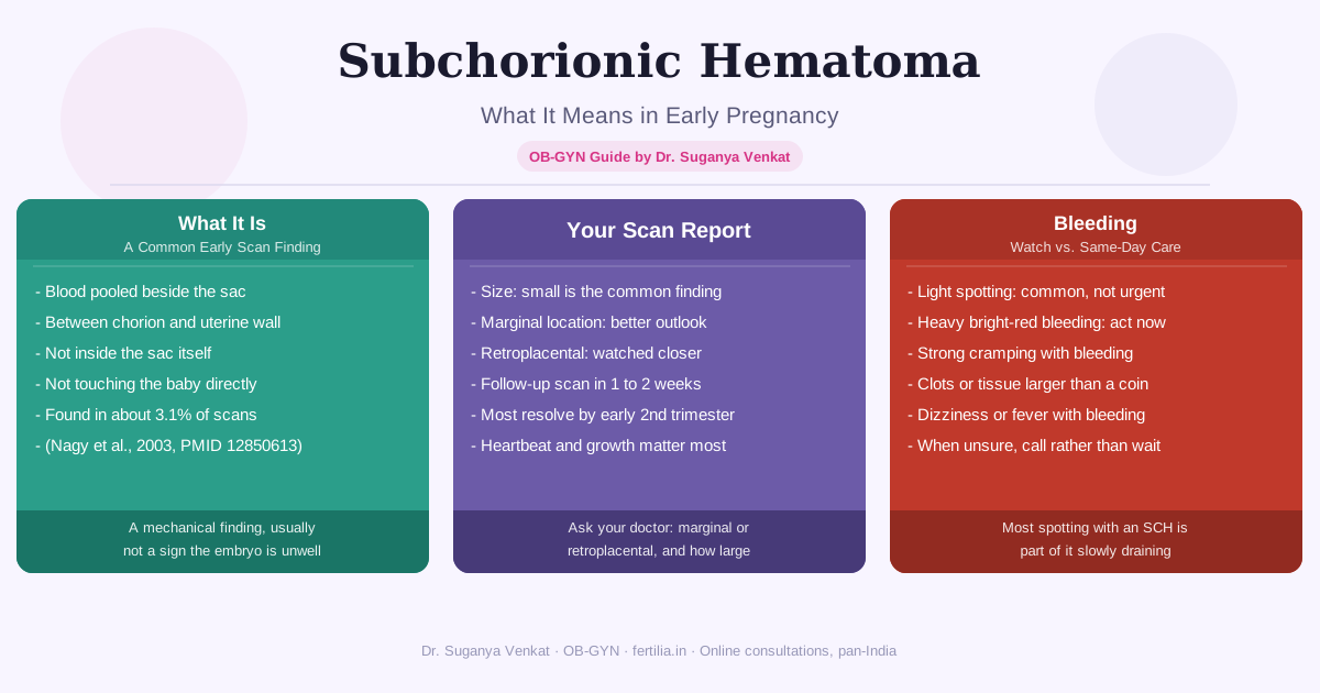
A subchorionic hematoma, sometimes written as subchorionic hemorrhage, is a collection of blood that has pooled between the chorion (the outer membrane surrounding the gestational sac) and the wall of the uterus. In simple terms, a small area at the edge of where the placenta is attaching has bled, and that blood has collected in a pocket rather than draining away immediately.
It is not blood inside the sac, and it is not blood touching your baby directly. It sits alongside the pregnancy, between the developing placenta and the uterine wall, which is exactly why it shows up as a dark, fluid-filled area on ultrasound rather than a change inside the sac itself.
This is a mechanical finding, not typically a sign that something is developmentally wrong with the embryo. The heartbeat, the growth, and the rest of the scan can all look completely normal alongside a subchorionic hematoma.
How common is this, and why does it show up now
A subchorionic hematoma is one of the most frequent incidental findings on a first-trimester scan. A large prospective study that compared 187 women with a first-trimester intrauterine hematoma against 6,488 women without one found the finding in 3.1% of a general obstetric population screened by routine early ultrasound (Nagy et al., 2003, Obstetrics & Gynecology, PMID 12850613). If you are seeing this on your own report, you are one of a meaningful number of women who see it, not an outlier.
It typically appears because early pregnancy scanning is now sensitive enough to pick up small collections of blood that, in earlier decades, would simply have gone unnoticed, sometimes passing as slightly heavier spotting or not being noticed at all. Finding it does not mean it is new or that something has just gone wrong. It often means the scan is doing exactly what it is meant to do: showing detail that was always too small to see before.
What your scan report is telling you
Two details on your report matter more than the word “hematoma” itself: size and location.
Size. Your report may give a measurement in millimetres, or describe the hematoma as small, moderate, or large relative to the gestational sac. A smaller hematoma, particularly one that is a small fraction of the sac’s own size, is the far more common finding and carries the better outlook. A larger one is watched more closely, but “larger” does not automatically mean the pregnancy is in danger. It means your doctor will want a repeat scan sooner to see whether it is growing, staying the same, or already resolving.
Location. This is the detail that matters clinically more than size alone. A marginal hematoma sits at the edge of the placenta, away from where the placenta is directly implanting into the uterine wall. A retroplacental hematoma sits directly behind the placenta itself. In the same study referenced above, a retroplacental position was the specific factor associated with a meaningfully higher risk of complications, while the presence or absence of bleeding symptoms did not change the outcome on its own (Nagy et al., 2003, PMID 12850613). If your report specifies “marginal,” that is the more reassuring of the two locations.
What this means in practice:
- Ask your doctor whether your hematoma is described as marginal or retroplacental, and roughly how large it is compared to the sac
- A small, marginal hematoma with a normal heartbeat is the most common combination, and it is the one with the best outlook
- Your doctor will schedule a follow-up scan, usually within one to two weeks, mainly to check whether it is shrinking
- Most hematomas that are going to resolve do so by the end of the first trimester or early in the second
If you have a subchorionic hematoma on your report and you are unsure what your specific measurements mean for you, I would rather you ask directly than sit with the uncertainty. WhatsApp +91 99402 70499.
What the evidence shows about outcomes
This is the part women searching for this term want most: does a subchorionic hematoma mean a higher chance of miscarriage.
The honest, evidence-based answer has two parts, and both matter.
Part one: a subchorionic hematoma is associated with a somewhat higher risk, on average, across large groups of women. A systematic review and meta-analysis pooling seven studies, covering 1,735 women with a subchorionic hematoma against 70,703 without one, found the rate of spontaneous pregnancy loss was 8.9% in women without a hematoma compared with 17.6% in women with one (pooled odds ratio 2.18) (Tuuli et al., 2011, Obstetrics & Gynecology, PMID 21508763). The same review found associations with preterm delivery and placental abruption as well.
Part two: this is a population-level average, and it means the large majority of individual pregnancies with a subchorionic hematoma continue without a pregnancy loss. The same data, read the other way, says that even in the group with a hematoma, more than 8 in 10 pregnancies did not end in loss. The “number needed to harm” in that review was 11 for spontaneous abortion, meaning the researchers estimated that for every 11 women diagnosed with a subchorionic hematoma, statistically one additional loss occurred compared with what would have happened without it. Ten of those eleven women’s pregnancies were unaffected by the hematoma at all.
I explain it to patients this way: a subchorionic hematoma is a real finding that shifts a probability, in the same direction that any complication of pregnancy shifts a probability, and it does not tell you which outcome is coming for you individually. What tells you more, week by week, is your own follow-up scan: is the heartbeat still there, is the pregnancy still growing on track, and is the hematoma itself shrinking. Those three answers matter far more than the word on the first report that found it. If you are also tracking your beta-hCG numbers alongside your scans, a normally rising level is another reassuring sign alongside the hematoma itself shrinking.
A note on size and risk together: larger hematomas and retroplacental hematomas carry a somewhat higher risk within this picture than small, marginal ones, which is why your doctor’s specific description of your finding is worth more than the general statistic. Ask about your own numbers rather than only the average.
Activity: what you can keep doing, and what to ease off
There is limited high-quality evidence that strict bed rest changes the outcome of a subchorionic hematoma, and complete bed rest carries its own downsides, including reduced circulation and unnecessary anxiety from being confined at home. What I usually recommend instead is a graded, common-sense approach rather than either extreme.
What is usually fine to continue:
- Gentle walking and normal daily movement around the house and for errands
- Light household activity
- Working from a desk, sitting, and normal day-to-day routines
What is usually asked to pause until the hematoma is smaller or resolved:
- Strenuous exercise, heavy lifting, and high-impact workouts
- Intercourse, until your doctor confirms the hematoma is stable or resolving (this is sometimes called pelvic rest)
- Long-haul travel, particularly if there is any active bleeding
- Tampon use, though this is rarely relevant this early in pregnancy
This is guidance to discuss with your own doctor, since the specific recommendation depends on your hematoma’s size, location, and whether you have any active bleeding at the time. For general guidance on what movement is safe trimester by trimester, our pregnancy exercise guide covers the broader picture alongside this. It is also worth saying plainly: a subchorionic hematoma is not something you caused through activity before it was found, and it is not something a woman needs to blame herself for managing “wrong” once it is diagnosed. Watchful monitoring, not fear, is what actually helps here.
The bleeding pattern that needs same-day care
Light spotting, brown discharge, or old-looking blood is a common accompaniment to a subchorionic hematoma as it slowly drains, and on its own it is not usually an emergency.
Contact your doctor the same day, or go to the nearest hospital, if you have:
- Heavy, bright red bleeding that soaks through a pad within an hour
- Bleeding accompanied by strong, cramping pain, similar to period pain or stronger
- Passage of tissue or clots larger than a coin
- Dizziness, fainting, or feeling unusually unwell alongside bleeding
- Fever alongside bleeding
Outside of these specific signs, if your bleeding is light and painless, it is reasonable to note it, mention it at your next visit or call, and continue your planned follow-up scan rather than treating every spot of blood as an emergency. Knowing the difference between “watch and mention” and “go now” is often what brings the most relief to women managing this finding through the following weeks.
If you are bleeding right now and unsure which category you’re in, please reach out rather than waiting it out alone: WhatsApp +91 99402 70499.
What is a subchorionic hematoma called in Hindi and Tamil?
| Language | Term | Meaning |
|---|---|---|
| Hindi | गर्भाशय में खून का थक्का (garbhashay mein khoon ka thakka) | Blood clot in the uterus |
| Hindi (as often searched) | pregnancy mein khoon ka clot | Blood clot in pregnancy |
| Tamil | கருப்பையில் இரத்தக் கட்டி (karuppaiyil iratha kaddi) | Blood clot in the uterus |
If your family or your report uses one of these terms rather than “subchorionic hematoma,” it is describing the same finding covered in this guide.
Frequently Asked Questions
Will a subchorionic hematoma cause a miscarriage?
Most do not. Large studies show the majority of pregnancies with a small or marginal subchorionic hematoma continue without a loss, and the finding is one factor among several your doctor weighs, alongside your heartbeat, growth, and follow-up scans, not a diagnosis of an outcome on its own.
How long does a subchorionic hematoma take to resolve?
This varies by size. Small hematomas often resolve within a few weeks, and most that are going to resolve do so by the end of the first trimester or early in the second trimester. Your doctor’s follow-up scan schedule is designed to track this specifically.
Can I still see my baby’s heartbeat if I have a subchorionic hematoma?
Yes. A subchorionic hematoma sits alongside the gestational sac, between the chorion and the uterine wall, not inside the sac where the embryo and heartbeat are seen. Many women with a hematoma have a completely normal heartbeat and growth on the same scan.
Do I need bed rest for a subchorionic hematoma?
Usually not strict bed rest. Most doctors recommend easing off strenuous exercise, heavy lifting, and intercourse until the hematoma is stable or smaller, while continuing normal gentle daily activity. Ask your own doctor for guidance specific to your hematoma’s size and location.
Is spotting normal with a subchorionic hematoma?
Light spotting or brown discharge is common as the hematoma slowly drains and is not usually an emergency on its own. Heavy bright red bleeding, strong cramping, passing clots or tissue, or bleeding with dizziness or fever needs same-day medical assessment.
Does the location of the hematoma matter?
Yes. A marginal hematoma, at the edge of the placenta, is generally associated with a better outlook than a retroplacental hematoma, which sits directly behind the placenta. Ask your doctor which location your report describes.
Will this happen again in a future pregnancy?
A subchorionic hematoma in one pregnancy does not mean it will recur in a future one. It is usually a localised, mechanical event in that specific pregnancy rather than a marker of an ongoing condition that needs pre-pregnancy investigation.
A subchorionic hematoma is one of the findings I most often need to talk women through calmly, because the word “hematoma” sounds far more alarming than what the finding usually turns out to mean. If you have this on your report and want to go through your specific scan together, reach out: WhatsApp +91 99402 70499. For more on what your early scans show week by week, see our early pregnancy scan guide, and our pregnancy resource guide covers what to expect and monitor through the rest of the first trimester.