If you have had a previous C-section and your scan report now mentions “placenta accreta” or “placenta accreta spectrum,” it is natural to feel your stomach drop. The word “accreta” is unfamiliar, the mention of possible hysterectomy is frightening, and a quick search online tends to surface the most severe cases rather than the full picture.
Here is what matters most before anything else: placenta accreta spectrum is a known, well-studied condition. When it is picked up early on a scan, and delivery is planned at a hospital with the right team and resources in place, outcomes for both mother and baby are good. The finding on your report is not a crisis unfolding. It is information that lets your care team prepare properly, well before your due date.
This guide explains what placenta accreta spectrum actually means, why prior C-sections are the main risk factor, how it is diagnosed, and how a safe delivery is planned around it.
What this post covers:
- What placenta accreta, increta, and percreta mean, and how they differ from placenta previa
- Why a prior C-section is the dominant risk factor, and how the risk changes with each surgery
- How it is diagnosed: ultrasound findings and when MRI is used
- What symptoms, if any, you might notice
- How delivery is planned, including the honest picture on hysterectomy
- How this connects to postpartum haemorrhage risk
What Placenta Accreta Spectrum Means
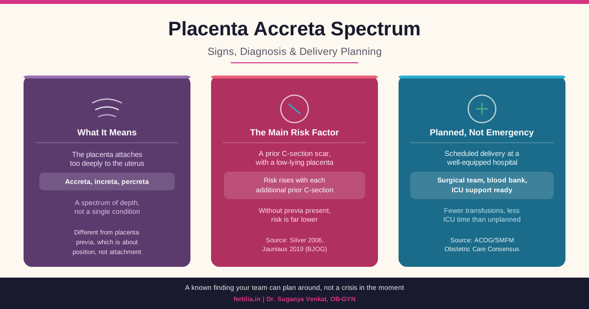
In a typical pregnancy, the placenta attaches to the lining of the uterus (the decidua) and stays there until it separates naturally after your baby is born. Placenta accreta spectrum (PAS) is the umbrella term for what happens when that attachment goes deeper than it should, because the decidua is thin, scarred, or missing in the area where the placenta implants (Jauniaux E et al., American Journal of Obstetrics and Gynecology, 2019, PMID 30716286).
There are three grades within the spectrum, and the terminology matters because it describes depth, not danger in a vague sense:
- Placenta accreta (sometimes called accreta vera): the placenta attaches directly to the surface of the muscle wall of the uterus (the myometrium) without the usual cushioning layer in between. This is the least deep form.
- Placenta increta: the placenta invades into the muscle wall itself.
- Placenta percreta: the placenta grows all the way through the muscle wall and can reach the outer surface of the uterus, and in some cases nearby structures such as the bladder.
A precise diagnosis of which of the three you have is usually only fully confirmed at delivery, sometimes with pathology review afterward. Antenatally, your team works from ultrasound findings that suggest PAS is present and roughly how extensive it appears, and plans your delivery around that picture.
Placenta previa, which we cover in detail in our dedicated guide, is a related but separate finding. Previa describes how close the placenta sits to the cervix. Accreta spectrum describes how deeply the placenta has grown into the uterine wall. The two often occur together, especially in women with a prior C-section, and your report may mention one, the other, or both.
Why a Prior C-Section Is the Main Risk Factor
The single biggest driver of placenta accreta spectrum is a scar from a previous C-section combined with a placenta that implants low, over or near that scar. A scar disrupts the normal lining of the uterus at that spot, and if the placenta happens to implant there in a later pregnancy, it can attach more deeply than it should, because the tissue it is meant to attach to is not fully intact.
The risk rises with each additional C-section, and it rises specifically in the presence of placenta previa. A landmark study that has shaped how this risk is counselled found that among women with placenta previa, the risk of placenta accreta was approximately 3% with one prior C-section, 11% with two, 40% with three, 61% with four, and 67% with five or more (Silver RM et al., Obstetrics & Gynecology, 2006, PMID 16738145). Without placenta previa present, the risk at any given number of prior C-sections is far lower. This is why your doctor pays close attention specifically when both factors, a scar and a low or previa placenta, are present together.
A prior C-section also raises the chance of placenta previa itself occurring in a later pregnancy, which is part of why the two conditions cluster together in women with multiple prior C-sections.
Other, less common contributors include any procedure that can scar the uterine lining: fibroid removal (myomectomy), dilatation and curettage (D&C), hysteroscopic surgery, endometrial ablation, or a previous C-section scar ectopic pregnancy. Advanced maternal age, IVF conception, and a short gap of under a year since your last C-section are additional factors that can raise the likelihood of a low placenta, which is the pathway through which accreta risk rises.
If none of these apply to you and your placenta is normally positioned, PAS is very unlikely. This is a condition that concentrates heavily in a specific, identifiable group of women: those with a prior C-section and a placenta that has implanted low or over the scar. If your scan does not flag a low-lying or previa placenta, this is not something you need to carry as background worry.
Scan report mention placenta accreta or a low placenta over your C-section scar? Dr. Suganya Venkat reviews your specific findings and talks you through what monitoring and delivery planning look like for your case. Chat on WhatsApp
How Placenta Accreta Spectrum Is Diagnosed
Ultrasound is the main diagnostic tool, and it is usually done by an experienced sonographer, sometimes including a transvaginal scan for a clearer view, particularly when the placenta sits at the back of the uterus. If you have had a prior C-section and this scan shows a low-lying or previa placenta, your team will specifically look for accreta spectrum signs at your 18 to 20 week anomaly scan, and follow up again later in pregnancy if anything is uncertain (Jauniaux E et al., BJOG, 2019, PMID 30260097).
What the sonographer is looking for on the scan:
- Irregular spaces within the placenta with unusual blood flow patterns, sometimes described as a “moth-eaten” or lacunar appearance
- Loss of the normal clear boundary layer between the placenta and the uterine muscle
- Thinning of the muscle wall beneath the placenta, particularly near a C-section scar
- Irregularity where the uterus meets the bladder
- Unusual blood vessels bridging the placenta and surrounding tissue
No single one of these findings confirms or rules out PAS on its own. Your radiologist or maternal-fetal medicine specialist puts the whole picture together, alongside your surgical history and where exactly the placenta sits, to reach a level of suspicion that guides how your delivery is planned.
MRI is not used routinely. When an experienced sonographer has done a thorough ultrasound, MRI usually does not add meaningful extra information, and current guidance does not recommend it as a routine step. It is used selectively: when the ultrasound view is technically difficult, when the placenta is at the back of the uterus and harder to assess, or when there is a specific question about how far invasion might extend toward the bladder that could change the surgical plan.
If your scan raises suspicion of PAS, early referral to a specialist maternal-fetal medicine team matters. This is not a diagnosis to sit with at a general clinic; the earlier a specialist team is involved, the more time there is to plan your delivery properly.
Symptoms: Often None Until Delivery
This is a point worth stating plainly, because it surprises many women: placenta accreta spectrum usually causes no symptoms during pregnancy. Most cases are identified entirely through routine ultrasound, not because a woman noticed something was wrong.
When symptoms do occur, the most notable one is painless vaginal bleeding in the second or third trimester, particularly in women who also have placenta previa. This overlaps with the bleeding pattern of previa itself, which is why the two conditions are assessed together. Any vaginal bleeding after 20 weeks needs same-day medical evaluation, regardless of whether PAS has been raised as a possibility.
The absence of symptoms is exactly why the scan matters so much. A woman can feel entirely well through pregnancy and still have a finding on ultrasound that changes how and where her delivery needs to happen.
How Delivery Is Planned
This is the part that tends to worry women the most, so let’s walk through it directly.
Delivery is planned in advance, at a hospital equipped for it. When PAS is suspected or confirmed antenatally, the safest approach is a scheduled C-section, timed before labour begins on its own, at a hospital with an experienced surgical team, a well-stocked blood bank ready for the possibility of significant blood loss, and intensive care support available if needed. Planning ahead like this, rather than dealing with PAS as an unexpected finding during an emergency delivery, is consistently associated with fewer blood transfusions, less need for intensive care, and a shorter hospital stay afterward (American College of Obstetricians and Gynecologists and Society for Maternal-Fetal Medicine, Obstetrics and Gynecology, 2018, PMID 30461695). This is the entire reason antenatal diagnosis through ultrasound matters as much as it does: it turns an unpredictable situation into a prepared one.
Timing is usually earlier than a standard C-section, often planned for somewhere in the mid-to-late 30s of weeks, balancing the risks of a slightly early delivery against the risk of going into labour unexpectedly with PAS present. If you go into labour on your own, or start bleeding, before your planned date, your team will act on that at whatever point you present.
The honest picture on hysterectomy. For many women with confirmed placenta accreta spectrum, especially the more invasive increta and percreta forms, the safest plan is to deliver the baby and then remove the uterus in the same surgery (a cesarean hysterectomy), without attempting to separate the placenta first. This is not because something has gone wrong. Attempting to peel away a deeply attached placenta is what causes the dangerous bleeding associated with PAS; leaving it in place and proceeding directly to hysterectomy is the deliberate, planned, safer choice in cases where invasion is extensive. Your surgical team will talk you through, before your delivery date, whether your specific scan findings point toward this being likely, so it is a known part of your plan rather than a decision made in the middle of an emergency.
In selected, less extensive cases, and with a specialist team experienced in it, more conservative approaches that aim to preserve the uterus are sometimes possible. This is an individual conversation to have with your maternal-fetal medicine specialist based on your specific scan findings, not something to assume either way from a general article.
What the surgical team has ready. For a planned PAS delivery, the operating team typically includes an experienced obstetric surgeon, anaesthesia, the blood bank on standby with cross-matched blood ready, and often a urologist on call in case the bladder is involved. This is exactly why delivery at a well-equipped hospital, arranged in advance, is the safest path: everything that might be needed is already in the room.
Planning delivery around a placenta accreta spectrum diagnosis? Dr. Suganya Venkat helps you understand your scan findings and coordinates the right level of hospital and team for your delivery. Chat on WhatsApp
The Connection to Postpartum Haemorrhage
Placenta accreta spectrum is one of the recognised causes of significant bleeding at delivery, which is exactly why the planning described above matters so much. Our complete guide to postpartum haemorrhage explains how hospitals prepare for and manage heavy bleeding after delivery in general. With PAS specifically, that preparation starts earlier, with blood products cross-matched and ready before your surgery even begins, precisely because the bleeding risk is anticipated rather than unexpected.
If you are also weighing a vaginal delivery after a prior C-section for a future pregnancy, it is worth knowing that PAS risk is one of the factors that shapes whether VBAC is a safe option to discuss with your doctor, since each additional C-section changes the accreta risk calculation for the pregnancy after that. For a full walkthrough of what a planned or emergency C-section itself involves, see our C-section delivery guide.
Frequently Asked Questions
What is placenta accreta spectrum?
It is a condition where the placenta attaches too deeply to the wall of the uterus instead of separating normally after delivery. It ranges from accreta (attached to the surface of the muscle wall) to increta (grown into the muscle) to percreta (grown all the way through it). It is most often related to a scar from a previous C-section combined with a low-lying placenta.
What are the symptoms of placenta accreta?
Most women with placenta accreta spectrum have no symptoms at all during pregnancy. It is usually found on a routine ultrasound. When symptoms do occur, painless vaginal bleeding in the second or third trimester is the most common one, particularly when placenta previa is also present.
Does having a C-section always mean I am at risk for placenta accreta in my next pregnancy?
No. The risk is specifically tied to a scar plus a placenta that implants low or over that scar in a later pregnancy. A single prior C-section with a normally positioned placenta carries a low risk. The risk rises meaningfully with each additional C-section, especially when placenta previa is also present.
How is placenta accreta diagnosed?
Mainly through ultrasound performed by an experienced sonographer, sometimes including a transvaginal scan, looking for specific signs like irregular blood-filled spaces in the placenta and thinning of the muscle wall underneath it. MRI is used selectively, not routinely, usually when the ultrasound view is technically difficult or the placenta is at the back of the uterus.
Will I need a hysterectomy if I have placenta accreta?
For more invasive forms, particularly increta and percreta, delivering the baby and removing the uterus in the same planned surgery is often the safest approach, because attempting to separate a deeply attached placenta is what causes dangerous bleeding. This is a planned decision discussed with you in advance, not an emergency measure. In some less extensive, carefully selected cases, uterus-preserving approaches may be possible with a specialist team; this is an individual conversation with your maternal-fetal medicine specialist.
Can placenta accreta be prevented?
There is no way to prevent it once a pregnancy has occurred, but the underlying risk can be reduced over time by keeping the number of C-sections as low as medically appropriate, and by discussing VBAC as an option in future pregnancies when it is safe to do so. If you already have a diagnosis in this pregnancy, the focus shifts from prevention to safe, planned delivery.
Where should I deliver if I have placenta accreta spectrum?
At a hospital with an experienced obstetric surgical team, a well-equipped blood bank, and intensive care support available, ideally one with specific experience managing PAS deliveries. If your local hospital does not have this level of resource, your doctor will usually recommend referral to a larger centre for your delivery, arranged well ahead of your due date.
The Bottom Line
A placenta accreta spectrum finding on your scan is exactly that: a finding, not a verdict. It tells your care team something important that they can plan around, well before you go into labour. The risk factors are well understood, the diagnostic signs are well described, and the safest path, a scheduled delivery at a properly equipped hospital with the right team assembled in advance, has consistently better outcomes than an unplanned one.
At Fertilia, Dr. Suganya Venkat has guided women through this exact conversation: reviewing the scan findings, explaining honestly what they mean, and coordinating with the right hospital and team so that delivery day is prepared for, not feared. If your scan has raised this possibility, the most useful next step is simply to talk it through with your obstetrician and confirm your delivery plan is in place.
Have a placenta accreta finding you want explained clearly? Dr. Suganya Venkat reviews your report and helps you understand your delivery plan, over a video consultation. Chat on WhatsApp
Dr. Suganya Venkat is an OB-GYN with a DNB from GKNM Hospital, Coimbatore, an MD Pathology from CMC Vellore, and 5 Gold Medals in MBBS from SRMC. She has 15+ years of clinical experience in obstetrics and women’s health.