Your gynaecologist writes “breast scan” on the referral, and when you call the diagnostic centre, the front desk asks: “Ultrasound or mammogram?” You had assumed those were the same thing. They are not. Each scan answers a different question, each has a situation where it is the better choice, and the reports they produce look different from each other. Understanding which one to do, and what to do with the numbers on the report, takes away a great deal of unnecessary anxiety.
For more on this, read our guide on Ovarian Cyst on Ultrasound. I am a radiologist, and breast imaging is a routine part of my week. You may have come across my guide to reading a follicular study report, which takes the same approach: translating the numbers on the page into something that makes sense. This guide does the same for breast scans. It explains what each scan is, why younger Indian women are often sent for ultrasound first, how to read the BI-RADS score on any breast report, and what the scans cost in India in 2026.
What this post covers:
- What a breast ultrasound does, and how it differs from a mammogram
- Why breast density matters, and why it changes which scan works better
- When each scan is the right first step
- Reading your breast scan report: BI-RADS categories in plain language
- What the scans cost at labs and hospitals in India
What a breast ultrasound is
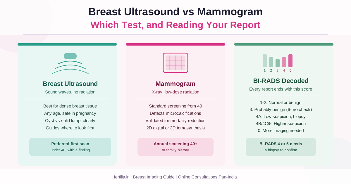
A breast ultrasound uses high-frequency sound waves to create a real-time image of the tissue inside the breast. There is no radiation, no compression of the breast, and no specific age restriction. The scan takes around 15 to 20 minutes. I move a handheld probe across the breast surface with a little gel and generate images on a screen as I go.
Ultrasound is very good at distinguishing a fluid-filled cyst from a solid lump. A simple cyst, which is round, has thin walls, and is filled entirely with clear fluid, appears clearly different from a solid mass, which has no fluid interior and defined or irregular margins. This is the main thing ultrasound does well: it separates the “this is definitely a cyst” finding from the “this is a solid mass that needs further assessment” finding, often at a single visit.
Ultrasound also performs well in dense breast tissue. This matters more than most women realise, and I will explain why in the section below.
What ultrasound does not show well: very fine specks of calcium called microcalcifications. These tiny calcium deposits, which can sometimes be an early sign of certain breast changes, are best visualised on mammography, not ultrasound. This is one of the reasons mammography remains the standard screening tool for women above a certain age.
What a mammogram is
A mammogram is an X-ray of the breast. The breast is placed on a plate, compressed gently (and briefly), and an image is taken from two angles for each side. The compression, while uncomfortable for a moment, is necessary to spread the tissue so structures within it are clearly visible.
There are two types. A standard 2D digital mammogram takes flat images from the front and the side. A 3D mammogram (tomosynthesis) takes multiple images at slightly different angles and produces a layered picture, similar in concept to a CT scan but for the breast. Tomosynthesis is better at finding cancers hidden behind overlapping tissue and is increasingly available in larger cities and cancer centres in India.
Mammography excels at detecting microcalcifications and is the most studied, most validated screening method we have for breast cancer in women above 40. It is the test that has the largest body of evidence behind it for reducing mortality when used as a routine screening tool.
Its limitation is that it depends on contrast. A lump shows up on mammography because it absorbs X-rays differently from the surrounding fat. When the breast tissue around it is also dense rather than fatty, that contrast disappears and a lump can hide in plain sight. This is the dense-breast problem.
Why breast density changes which scan works better
Breast density is not about how the breast feels to the touch. It is a description of how much fibroglandular tissue (gland and connective tissue) the breast contains compared to fat. Radiologists describe density in four categories, from almost entirely fatty to extremely dense.
On a mammogram, fat appears dark and fibroglandular tissue appears white. A dense breast has a lot of white. A small mass, which also appears white, can be invisible against a white background. A 2002 study published in Radiology that compared mammography, physical examination, and ultrasound across 27,825 patient evaluations found that in women with extremely dense breasts, mammogram sensitivity dropped to around 47 to 64 percent, compared to 97 percent in women with fatty breasts (Kolb TM et al., Radiology, PMID 12355001).
Ultrasound does not have this problem. Sound waves create contrast based on how tissue transmits and reflects them, not on X-ray density. A solid mass in a dense breast shows up on ultrasound in a way it might not on mammography. A landmark ACRIN 6666 study published in JAMA found that adding ultrasound to mammography in women with dense breasts and elevated risk found additional cancers that mammography alone had missed (Berg WA et al., JAMA 2008, PMID 18477782). A later Connecticut study confirmed similar additional cancer detection rates when supplemental ultrasound was offered to women with dense breasts in a general screening population (Hooley RJ et al., Radiology 2012, PMID 22723501).
For more on this, read our guide on Endometrial Cancer. Most Indian women under 40 have dense breasts by anatomy. This is why your doctor often starts with an ultrasound rather than a mammogram if you are in your 20s or 30s. It is not that mammography is wrong, it is that ultrasound tends to see more in this tissue type.
Which scan to do, and when
The choice is not always either/or. In practice, the two scans are often complementary.
Start with ultrasound if:
- You are under 40
- You have a specific finding: a lump you or your doctor can feel, breast pain localised to one spot, a nipple discharge
- You are pregnant or breastfeeding (no radiation)
For more on this, read our guide on Breast Lump & Self-Exam.
- A previous mammogram was assessed as dense and the report recommended supplemental ultrasound
- You are having a routine check at a diagnostic centre and your doctor has not specified which scan
Start with mammography if:
- You are 40 or older and due for routine annual screening
- Your doctor is looking specifically for microcalcifications
- You have a family history of breast cancer and your doctor has recommended mammographic screening
- You are following up a previous mammogram finding
Both together if:
- A lump has been found and needs full characterisation
- You are at elevated risk (family history, prior biopsy showing high-risk features) and your centre follows a combined protocol
- One scan raises a question the other is better placed to answer
If you are unsure, the simplest thing to do is tell the radiologist your age, your specific symptom or worry, and whether this is a routine check or a follow-up to something. We will advise you on the spot.
Unsure which scan applies to your situation, or have a report you want to understand? Dr. Suganya Venkat’s team offers online consultations across India. WhatsApp 919940270499 to book a ₹399 video consultation.
Reading your breast scan report: BI-RADS explained
Whether the report is from an ultrasound or a mammogram, it will end with a BI-RADS assessment category. BI-RADS stands for Breast Imaging Reporting and Data System and is published by the American College of Radiology. Every radiologist worldwide uses this system. Understanding it takes away most of the fear that comes from reading a report.
For more on this, read our guide on Thyroid Nodule on Ultrasound. There are seven categories.
BI-RADS 0: Incomplete. The current images are not enough to make a final assessment. Additional imaging is needed, either a different view on the same scan, a comparison with an older scan, or the other type of scan altogether. A BI-RADS 0 on an ultrasound often means a mammogram has been requested. A BI-RADS 0 on a mammogram often means an ultrasound of a specific area is needed. It is not a sign that something is wrong; it is a sign that more information is needed before a conclusion can be reached.
BI-RADS 1: Negative. Nothing has been found. The scan is normal. Routine screening applies.
BI-RADS 2: Benign. Something has been found and identified as definitely benign. Common examples include a simple cyst (clearly fluid-filled, round, smooth walls), a calcified fibroadenoma (a common benign lump that has calcified over time), or a fat-containing lesion. These findings are documented so future scans can note whether anything has changed, but they require no follow-up action. Many routine scans come back BI-RADS 2.
BI-RADS 3: Probably benign. Something has been found that has more than a 98 percent chance of being benign. The standard recommendation is a short-interval follow-up scan in six months to confirm it has not changed. Most BI-RADS 3 findings are oval solid nodules with smooth margins, likely fibroadenomas, which are very common in younger women. The six-month scan is to demonstrate stability, not because there is serious concern.
BI-RADS 4: Suspicious. This is the category that triggers the most worry and deserves the most explanation. A BI-RADS 4 finding has features that make it worth investigating, and a biopsy is usually recommended. The category is divided into three sub-levels:
- 4A (Low suspicion): The risk of malignancy is roughly 2 to 10 percent. Most 4A findings turn out to be benign at biopsy. Examples include a slightly irregular cyst wall or a solid nodule that does not quite fit the typical fibroadenoma pattern.
- 4B (Intermediate suspicion): The risk is approximately 10 to 50 percent. Tissue sampling is standard.
- 4C (Moderate concern): The risk is approximately 50 to 95 percent. There are features that raise more concern, though the finding does not tick every box for a BI-RADS 5.
BI-RADS 5: Highly suggestive of malignancy. The features seen have a greater than 95 percent likelihood of being malignant. A biopsy is necessary and prompt.
BI-RADS 6: Known biopsy-proven malignancy. This category is used when a scan is done on a lesion that has already been confirmed as cancerous by biopsy, usually to plan or monitor treatment.
For more on this, read our guide on Transvaginal Scan (TVS). A few things worth knowing about BI-RADS:
A BI-RADS 3, 4, or 5 does not mean you have cancer. It means the finding needs further assessment. The categories are a triage system, not a diagnosis. The actual diagnosis comes from a biopsy if one is needed.
The BI-RADS system is designed so that a 4A biopsy, if it comes back benign, is a success. It means the system worked and reassurance was obtained with evidence. Most 4A biopsies are benign.
If your report ends in BI-RADS 1, 2, or 3, there is nothing urgent to act on beyond following the follow-up schedule it recommends. If your report ends in BI-RADS 4 or 5, the next step is a tissue sample, arranged through your gynaecologist or a breast surgeon.
What breast scans cost in India in 2026
Prices vary by city, centre type, and whether the scan is unilateral (one breast) or bilateral (both sides). These are verified 2026 ranges based on published lab and hospital rates.
Breast ultrasound (bilateral):
- Diagnostic labs (Dr. Lal PathLabs, SRL, Metropolis): Rs 960 to 2,460
- Private hospitals and radiology centres: Rs 1,500 to 3,500
- Government hospitals: lower, vary by state
Digital mammogram (bilateral):
- Diagnostic labs and radiology centres: Rs 1,000 to 3,500
- CGHS beneficiaries: Rs 1,375 (metro/NABH rate)
- Government hospitals: nominal or free for women in many states
3D mammogram / tomosynthesis:
- Private hospitals and cancer centres: Rs 3,000 to 7,000
- Not available at most standalone diagnostic labs
Prices typically include the scan and a radiologist’s written report. If a consultation with the radiologist or a separate reporting fee is charged, ask about this in advance. Prices at government hospitals and under CGHS rates are significantly lower. Many major government cancer centres (Tata Memorial Mumbai, Kidwai Bengaluru, AIIMS Delhi, Adyar Cancer Institute Chennai) offer lower-cost mammography for women who meet their programme criteria.
Have a breast scan report you would like someone to review, or need to understand which scan makes sense for you? Dr. Suganya Venkat’s team is available online, pan-India, for a ₹399 video consultation. WhatsApp 919940270499 to get started.
Practical steps
If you have been referred for a breast scan and are not sure which one:
- Tell the centre your age. If you are under 40 and the referral does not specify, ultrasound is usually the starting point.
- Mention any symptoms you have: a lump, pain, nipple change, discharge.
- Bring any earlier scan reports if you have them. A comparison to a previous image often saves a follow-up visit.
If you have received a report and are not sure what to do with it:
- Find the BI-RADS number. That is the key line.
- BI-RADS 1 or 2: routine screening, no action required beyond the next scheduled scan.
- BI-RADS 3: book the six-month follow-up. Do not wait longer than recommended.
- BI-RADS 4 or 5: arrange a biopsy referral through your gynaecologist or surgeon. Do not delay, but do not panic either: even BI-RADS 5 findings require a tissue sample before any diagnosis is made.
If your report says “dense breast tissue” or notes that mammographic sensitivity is reduced: Ask whether a supplemental ultrasound is recommended. This is a routine, standard add-on in many centres. The mammogram and ultrasound together give a more complete picture than either alone when density is high.
For anything related to the breast findings and their implications for your fertility, your menstrual health, or your overall women’s health picture, your gynaecologist is the person to speak with. The radiology report describes what is in the image; your gynaecologist puts it in the context of your full health history.
For the broader picture of women’s health screening, the cervical cancer screening guide and the preconception checkup resource cover what other routine tests are recommended for women in their 20s, 30s, and 40s. If you have received other scan reports from a gynaecology workup, the guides to reading an antral follicle count report and reading an HSG report use the same plain-language approach.
Frequently asked questions
At what age should I start mammogram screening in India?
Most guidelines recommend starting annual mammography at 40 for women at average risk. The Indian Council of Medical Research and the Federation of Obstetric and Gynaecological Societies of India both recommend starting by 40, with some guidelines suggesting that women with a first-degree family history of breast cancer begin ten years earlier than the age at which the relative was diagnosed. Below 40, ultrasound is typically the preferred imaging method because of breast density.
What does “dense breasts” mean on my mammogram report?
Breast density describes how much glandular and connective tissue your breast contains relative to fat. Radiologists classify it into four categories. At the denser end, mammographic sensitivity drops because the tissue being looked at (fibroglandular) appears similar in colour on X-ray to a potential mass. This does not mean anything is wrong. It is a normal anatomical variation, more common in younger women. When a mammogram report notes high density, a supplemental ultrasound is often recommended to improve overall detection.
Can I do breast ultrasound instead of mammogram at 40?
For symptomatic women (a lump, pain, discharge), ultrasound is often the first scan at any age. For routine screening in women above 40, mammography is the standard because it detects microcalcifications, which ultrasound misses. In women above 40 with dense breasts, many breast radiologists recommend both together rather than one or the other. Your gynaecologist or the radiologist at your centre will advise based on your full picture.
My report says BI-RADS 3, do I need a biopsy?
No, not usually. BI-RADS 3 means the finding has a greater than 98 percent chance of being benign. The recommendation is a follow-up scan in six months to confirm stability. If the finding has not changed at six months, it is typically reclassified to BI-RADS 2. A biopsy for a BI-RADS 3 is considered only in specific circumstances, such as when a patient is about to undergo fertility treatment or surgery where a delay in the follow-up is not practical.
My report says BI-RADS 4A. How worried should I be?
BI-RADS 4A carries a risk of malignancy in the range of 2 to 10 percent. That means the large majority of 4A biopsies come back benign. A biopsy is recommended to find out definitively, not because cancer is likely, but because uncertainty is not useful for you or your doctor. Most 4A findings turn out to be fibroadenomas, benign cysts, or fibrocystic tissue. Get the biopsy done and act on the result.
How long does a breast ultrasound take, and does it hurt?
A bilateral breast ultrasound typically takes 15 to 25 minutes. There is no compression, no radiation, and no preparation required beyond removing upper-body clothing. The gel used is cool. The scan is not painful. Most women find it comfortable throughout.
Is there a breast ultrasound vs mammogram difference in detecting cancer?
The two scans are complementary rather than competitive. Mammography is more sensitive for microcalcifications and remains the most validated screening tool for population-level mortality reduction. Ultrasound is more sensitive in dense breast tissue and in characterising a specific lump or mass. When used together in the right situation, they are more useful than either alone. The choice of which to start with depends on your age, your breast density, whether you have a specific symptom, and what your doctor is looking for.
Dr. Rajashree NS is a Consultant Radiologist (MD Radio-diagnosis). At Fertilia she consults online, by video call, with women across India.
Evidence cited: Kolb TM et al., Radiology 2002 (PMID 12355001); Berg WA et al., JAMA 2008 (PMID 18477782); Hooley RJ et al., Radiology 2012 (PMID 22723501). BI-RADS categories: ACR BI-RADS Atlas 5th Edition, 2013.